

Clinical Features of Anthroponotic Cutaneous Leishmaniasis in a Major Focus, Southeastern Iran, 1994-2014
- PMID: 29317879
- PMCID: PMC5756304
Clinical Features of Anthroponotic Cutaneous Leishmaniasis in a Major Focus, Southeastern Iran, 1994-2014
Abstract
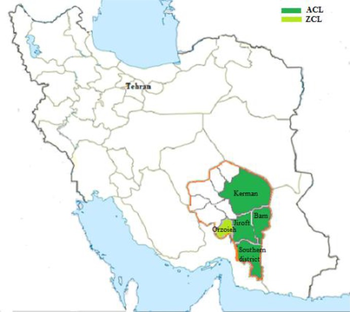
Background: Cutaneous leishmaniasis (CL) is associated with a broad and complex clinical spectrum of diseases. The objectives of this study were to assess the clinical features and identification of the causative agents of CL in a well-known focus of anthroponotic CL (ACL) caused by Leishmania tropica, southeast Iran.
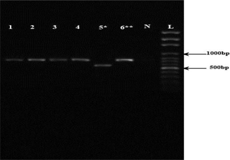
Methods: This study was performed randomly as a descriptive cross-sectional survey to evaluate 2000 CL patients by active and passive case-detection approaches in Kerman Province from 1994 to 2014. The ACL patients were confirmed by direct smear and 600 cases by one or a combination of intrinsic methods.
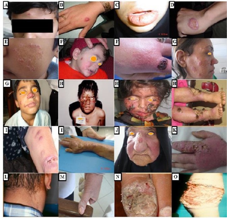
Results: Children aged <10 yr old were the most infected patients (P<0.001). The majority of the CL lesions were located in hands (46.3%), face (34.1%), legs (14.3%), and other parts of the body (5.3%). The mean number of lesions was 1.5 and most of the patients had single lesion (65%).Typical clinical lesions included papule (36.8%), followed by ulcerated nodule (20.7%), plaque (18.4%), and ulcerated plaque (18.5%). While among atypical clinical features, leishmaniasis recidivans (LR) (4.7%) and leishmanid (0.3%) were the dominant forms, followed by diffuse, disseminated, sporotrichoid, and erysipeloid types, 0.1% each, and then lymphedematous, lymphadenic, hyperkeratotic, paronychial, and mutilating types, 0.05% each. Based on various intrinsic methods the parasites isolated from the lesions were characterized as L. tropica.
Conclusion: ACL due to L. tropica presents numerous cases of localized form and diverse uncommon clinical presentations, which mimic other disease conditions. Therefore, physicians should be aware of such manifestations for selecting appropriate treatment modality.
Keywords: Clinical features; Cutaneous leismaniasis; Iran; Leishmania tropica.
Conflict of interest statement
Conflict of Interests The authors declare that there is no conflict of interests.
Figures
References
-
- Desjeux P. Leishmaniasis: current situation and new perspectives. Comp Immunol Microbiol Infect Dis. 2004;27(5):305–18. - PubMed
-
- Postigo JA. Leishmaniasis in the World Health Organization Eastern Mediterranean Region. Int J Antimicrob Agents. 2010;36 Suppl 1:S62–5. - PubMed
-
- Shirzadi MR, Esfahania SB, Mohebali M, et al. Epidemiological status of leishmaniasis in the Islamic Republic of Iran, 1983–2012. East Mediterr Health J. 2015;21(10):736–42. - PubMed
-
- Reithinger R, Dujardin JC, Louzir H, et al. Cutaneous leishmaniasis (Review). Lancet Infect Dis. 2007;7(9):581–96. - PubMed
LinkOut - more resources
Full Text Sources