

Feline dry eye syndrome of presumed neurogenic origin: a case report
- PMID: 29318025
- PMCID: PMC5753927
- DOI: 10.1177/2055116917746786
Feline dry eye syndrome of presumed neurogenic origin: a case report
Abstract
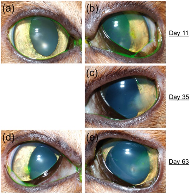
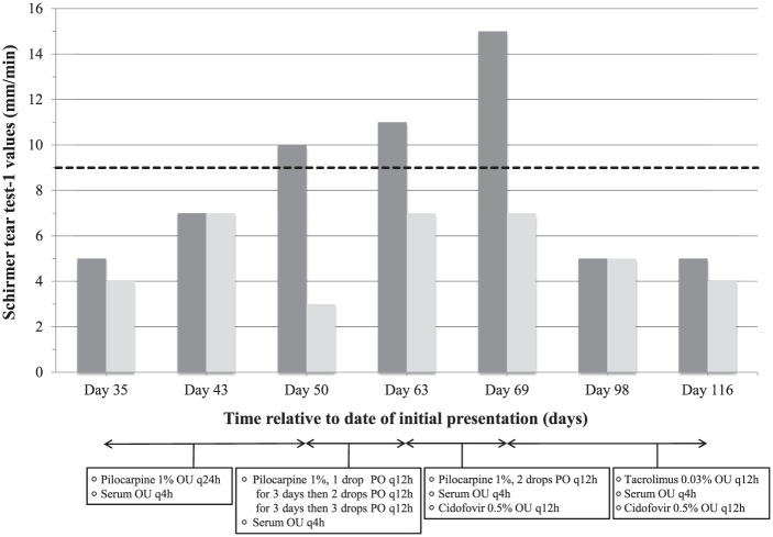
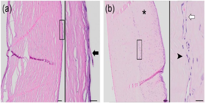
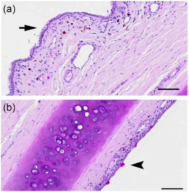
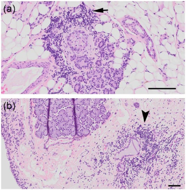
Case summary: A 14-year-old female spayed Abyssinian cat, which about 1 year previously underwent thoracic limb amputation, radiotherapy and chemotherapy for an incompletely excised vaccine-related fibrosarcoma, was presented for evaluation of corneal opacity in the left eye (OS). The ocular surface of both eyes (OU) had a lackluster appearance and there was a stromal corneal ulcer OS. Results of corneal aesthesiometry, Schirmer tear test-1 (STT-1) and tear film breakup time revealed corneal hypoesthesia, and quantitative and qualitative tear film deficiency OU. Noxious olfactory stimulation caused increased lacrimation relative to standard STT-1 values suggesting an intact nasolacrimal reflex. Various lacrimostimulants were administered in succession; namely, 1% pilocarpine administered topically (15 days) or orally (19 days), and topically applied 0.03% tacrolimus (47 days). Pilocarpine, especially when given orally, was associated with notable increases in STT-1 values, but corneal ulceration remained/recurred regardless of administration route, and oral pilocarpine resulted in gastrointestinal upset. Tacrolimus was not effective. After 93 days, the cat became weak and lame and a low thyroxine concentration was detected in serum. The cat was euthanized and a necropsy performed. Both lacrimal glands were histologically normal, but chronic neutrophilic keratitis and reduced conjunctival goblet cell density were noted OU.
Relevance and novel information: The final diagnosis was dry eye syndrome (DES) of presumed neurogenic origin, associated with corneal hypoesthesia. This report reinforces the importance of conducting tearfilm testing in cats with ocular surface disease, as clinical signs of DES were different from those described in dogs.
Conflict of interest statement
Conflict of interest: The authors declared no potential conflicts of interest with respect to the research, authorship, and/or publication of this article.
Figures
References
-
- Miller PE. Lacrimal system. In: Maggs DJ, Miller PE, Ofri R. (eds). Slatter's fundamentals of veterinary ophthalmology. 5th ed. St Louis, MO: Elsevier Saunders, 2013, pp 165–183.
-
- Matheis FL, Walser-Reinhardt L, Spiess BM. Canine neurogenic keratoconjunctivitis sicca: 11 cases (2006–2010). Vet Ophthalmol 2012; 15: 288–290. - PubMed
-
- Wagner F, Meyer-Lindenberg A, Heider HJ, et al. A comparison of corneal sensitivity between healthy cats and cats with corneal sequestra. Berl Munch Tierarztl Wochenschr 2003; 116: 427–431. - PubMed
-
- Zilstorff-Pedersen K. Quantitative measurements of the nasolacrimal reflex. Arch Otolaryngol 1965; 81: 457–462. - PubMed
Publication types
LinkOut - more resources
Full Text Sources
Other Literature Sources
Miscellaneous