Review
. 2017 Oct 4;2(3):176-191.
doi: 10.1016/j.synbio.2017.09.003. eCollection 2017 Sep.

Transcription control engineering and applications in synthetic biology

Affiliations
Review

Transcription control engineering and applications in synthetic biology

Michael D Engstrom et al. Synth Syst Biotechnol. .

Abstract

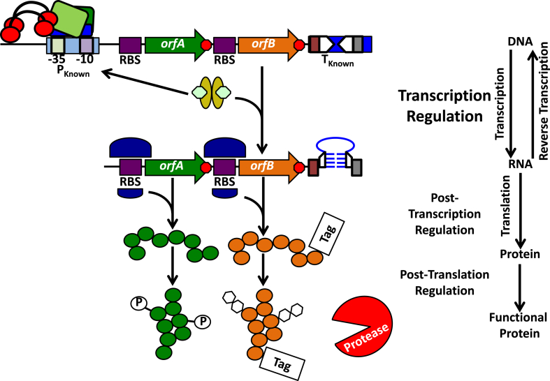
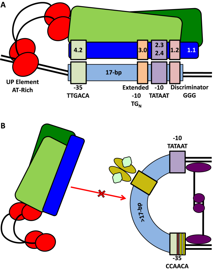
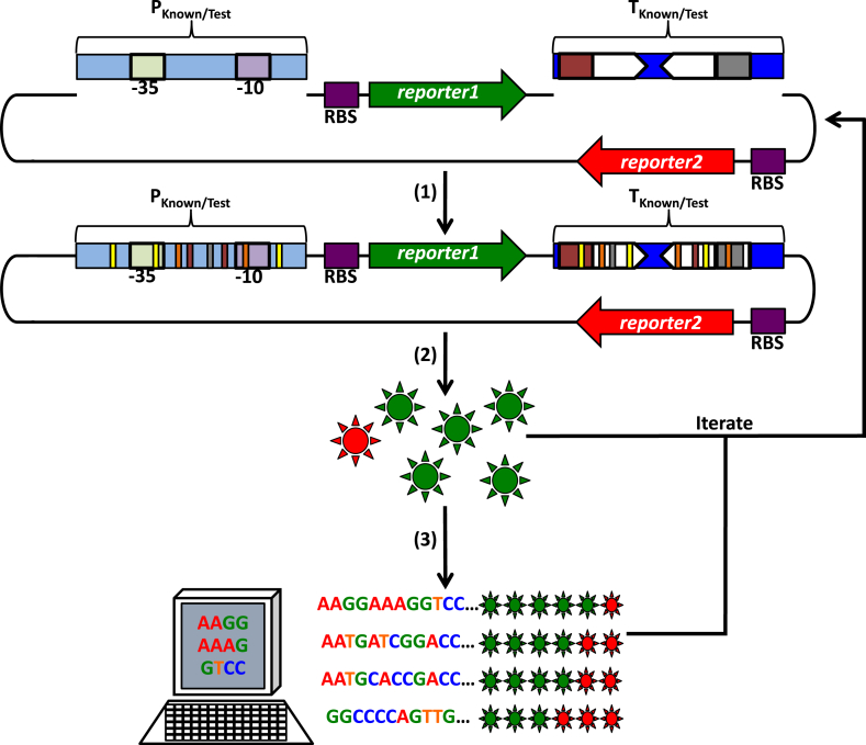
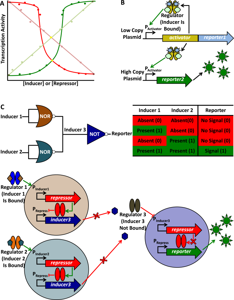
In synthetic biology, researchers assemble biological components in new ways to produce systems with practical applications. One of these practical applications is control of the flow of genetic information (from nucleic acid to protein), a.k.a. gene regulation. Regulation is critical for optimizing protein (and therefore activity) levels and the subsequent levels of metabolites and other cellular properties. The central dogma of molecular biology posits that information flow commences with transcription, and accordingly, regulatory tools targeting transcription have received the most attention in synthetic biology. In this mini-review, we highlight many past successes and summarize the lessons learned in developing tools for controlling transcription. In particular, we focus on engineering studies where promoters and transcription terminators (cis-factors) were directly engineered and/or isolated from DNA libraries. We also review several well-characterized transcription regulators (trans-factors), giving examples of how cis- and trans-acting factors have been combined to create digital and analogue switches for regulating transcription in response to various signals. Last, we provide examples of how engineered transcription control systems have been used in metabolic engineering and more complicated genetic circuits. While most of our mini-review focuses on the well-characterized bacterium Escherichia coli, we also provide several examples of the use of transcription control engineering in non-model organisms. Similar approaches have been applied outside the bacterial kingdom indicating that the lessons learned from bacterial studies may be generalized for other organisms.

Keywords: Bacteria; Metabolic engineering; Promoter; Synthetic biology; Termination; Transcription.


Figures

Fig. 1
Fig. 1
Overview of the central dogma of molecular biology. An operon encoding orfA and orfB is under the control of the indicated promoter (PKnown), which is engaged by the RNA polymerase holoenzyme (green and blue boxes, red circles, and black arcs). Sequences that will be transcribed into the ribosome binding sites (RBSs) for orfA and orfB (purple boxes) and the transcription terminator (TKnown; two inverted repeats [inverted arrowheads] flanked by a poly A-tract [red box] and poly U-tract [grey box]) are indicated. Red octagons represent translation stop codons. At the level of transcription regulation, alterations to the promoter and terminator sequences, the presence of transcription activators and repressors (depicted as golden ovals with green hexagon ligands bound), and nucleoid/supercoiling states may all influence orfA and orfB transcription (not shown). Through the process of transcription itself, the associated orfA and orfB mRNA is produced, which may in turn be bound by ribosomes (depicted as two variable size purple half circles) at the RBS. To terminate transcription, the depicted terminator sequence forms a stem-loop (blue), mediated by base pairing/stacking with the complementary sequences of the former inverted repeats (half arrowheads), and mRNA synthesis ceases at this site (poly A- and ploy U-tracts are indicated as above). Modifying RBS strength, RNA riboswitches, aptazymes (aptamer ribozymes), and RNA stability may each contribute to post-transcription regulation (not shown). Folding of a translated protein (green and orange circles) with the assistance of folding chaperones (not shown), enzymes that promote post-translation modifications such as phosphorylation (P in a white circle) or glycosylation (white hexagons), the presence of proteolytic enzymes (indicated in the figure and potentially targeted to a protein through a specific motif [Tag]) represent methods of post-translation regulation.
Fig. 2
Fig. 2
A representation of RNA polymerase holoenzyme-promoter interactions. (A) RNAP β and β′ subunits are indicated by green boxes. The RNAP α subunits (red circles and black arcs) are in contact with the bent upstream UP element. σ70 (dark blue box) sub-region 4.2 (pale green), 3.0 (pale orange), 2.3/2.4 (pale purple), 1.2 (pale pink), and 1.1 are associated with the indicated −35 sequence (pale green), extended −10 sequence (pale orange), −10 sequence (pale purple), discriminator (pale pink), and double stranded DNA, respectively. In this case, promoter elements are separated by 17-bp. (B) RNAP holoenzyme promoter binding is obstructed (red arrow and a red cross) by DNA bending mediated by nucleoid structuring factors or transcription factors (purple ovals, circles, and lines), other transcription regulators (golden ovals with green hexagon ligands bound) bound to operator sequences (gold box), spacing greater than 17-bp between the −35 and −10 sequences, and a −35 sequence deviating from the canonical −35 sequence (yellow and red bars). These figures are modified from the work by Browning and Busby .
Fig. 3
Fig. 3
A procedure to generate promoter and terminator libraries. (1) To test putative promoters as sites of transcription initiation, known promoters (PKnown) or random sequences (PTest) may be inserted upstream of a previously promoterless ribosome binding site (RBS)-reporter1 gene translational fusion (purple box with a green arrow). These promoters may be constitutive (shown) or subject to transcription regulation by activators and repressors (not shown). Pale green and pale purple boxes indicate the −35 and −10 promoter sequences, respectively. To test putative transcription terminators, known terminators (TKnown) or random sequences (TTest) may be inserted upstream of a promoterless RBS-reporter2 translational fusion (purple box with a red arrow) and downstream of the PKnown/Test-reporter1 transcription fusion above. Additional known or random terminators may be inserted in tandem, and a known terminator may be inserted downstream of reporter2 to ensure termination of transcription after RNAP reads-through this gene (not shown). Inverted white arrows represent the sequence forming a terminator stem-loop (inverted repeats), and red and grey boxes represent poly A- and poly U-tracts, respectively. (2) To introduce variability in promoter and terminator libraries (thin yellow, orange, red, grey, and white bars) these cis-regulatory elements may be derived from randomly sheared DNA, synthesized on demand with desired sequences, subjected to mutagenic PCR (polymerase chain reaction), or other similar methods. These cis-regulatory element constructs may be introduced into an organism of interest, reporter gene expression may be assessed for each library member (green and red suns), and this procedure may be iterated (starting with new random/mutated sequences or using library member sequences as inputs for further sequence variation generation) to achieve a desired result or a wider expression variability profile. (3) To identify sequence characteristics that may be responsible for reporter gene expression level differences (rows of green [gene expression from promoters] and red [gene expression from transcription read-through] suns) among library member sequences (indicated), these constructs should be sequenced and compared with other members or known promoter sequences (iterated as desired).
Fig. 4
Fig. 4
An overview of transcription regulation following analogue and digital expression dynamics. (A) A hypothetical transcription activity dynamics plot is depicted. Faint green and red lines represent transcription activity (activation and repression, respectively) subject to linear/analogue dynamics. Dark green and red curves represent activation and repression subject to step-change/digital dynamics, respectively. Individual points represent one hypothetical measurement of transcription activity. Concentrations of hypothetical activator or repressor ligands increase from left to right on the horizontal axis, and transcription activity increases from the bottom to the top of the vertical axis. (B) An example of a system subject to linear/analogue dynamics. This model is modified from the work of Daniel et al. . Activator (gold ovals) fused to Reporter 1 (blue suns) is produced from a gene expressed from a low copy plasmid and subject to a positive feedback (auto-activation) loop at PActivator (green arrow). Activator (bound to inducer) also binds target sites on a high copy number plasmid activating reporter2, again at PActivator, and simultaneously titrates Activator from the low copy plasmid above. Based on inducer concentration, this model allows for log-linear titration of Reporter 2 (green suns) levels . (C) A digital AND logic gate (engineered from two NOR gates and a NOT gate) is symbolically represented along with the associated truth table. The depicted model is modified from the work of Tamsir et al. . This circuit is distributed among three co-cultured cells (pale brown, blue, and purple circles), and the scenario depicted represents the ON output state, when Reporter (green suns) is produced (1 and a green box in the truth table). Both inducer 1 (brown diamond) and inducer 2 (blue pentagon) are present (1 and a green box in the truth table) and bound to Regulators 1 and 2, respectively. These regulators bind PInducer1 and PInducer2, respectively, activating repressor (green arrow). Repressor is subsequently produced and represses inducer3 (red crossbar), the gene encoding the producer of inducer 3 (purple hexagon), at PRepress. Inducer 3 is not secreted (red arrow with a cross), and Regulator 3 (without inducer 3 bound) binds PInducer3 repressing repressor. Subsequently, Repressor does not repress reporter, at PRepress, leading to reporter expression and Reporter production. In the absence of either or both inducers 1 and 2 (0 and red boxes in the truth table) inducer3 is not repressed, which leads to inducer 3 secretion. Regulator 3 (with inducer 3 bound) activates repressor, at PRepress. Repressor subsequently represses reporter, blocking Reporter production (0 and a red box in the truth table).

References

    1. Crick F. Central dogma of molecular biology. Nature. 1970;227:561–563. - PubMed
    1. Cech T.R. Structural biology. The ribosome is a ribozyme. Science. 2000;289:878–879. - PubMed
    1. Ban N., Nissen P., Hansen J., Moore P.B., Steitz T.A. The complete atomic structure of the large ribosomal subunit at 2.4 A resolution. Science. 2000;289:905–920. - PubMed
    1. Nissen P., Hansen J., Ban N., Moore P.B., Steitz T.A. The structural basis of ribosome activity in peptide bond synthesis. Science. 2000;289:920–930. - PubMed
    1. Muth G.W., Ortoleva-Donnelly L., Strobel S.A. A single adenosine with a neutral pKa in the ribosomal peptidyl transferase center. Science. 2000;289:947–950. - PubMed