. 2018 Mar;28(2):173-182.
doi: 10.1111/jon.12485. Epub 2018 Jan 10.

Image Registration to Compensate for EPI Distortion in Patients with Brain Tumors: An Evaluation of Tract-Specific Effects

Affiliations

Image Registration to Compensate for EPI Distortion in Patients with Brain Tumors: An Evaluation of Tract-Specific Effects

Angela Albi et al. J Neuroimaging. 2018 Mar.

Abstract

Background and purpose: Diffusion magnetic resonance imaging (dMRI) provides preoperative maps of neurosurgical patients' white matter tracts, but these maps suffer from echo-planar imaging (EPI) distortions caused by magnetic field inhomogeneities. In clinical neurosurgical planning, these distortions are generally not corrected and thus contribute to the uncertainty of fiber tracking. Multiple image processing pipelines have been proposed for image-registration-based EPI distortion correction in healthy subjects. In this article, we perform the first comparison of such pipelines in neurosurgical patient data.

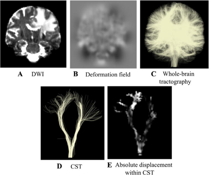
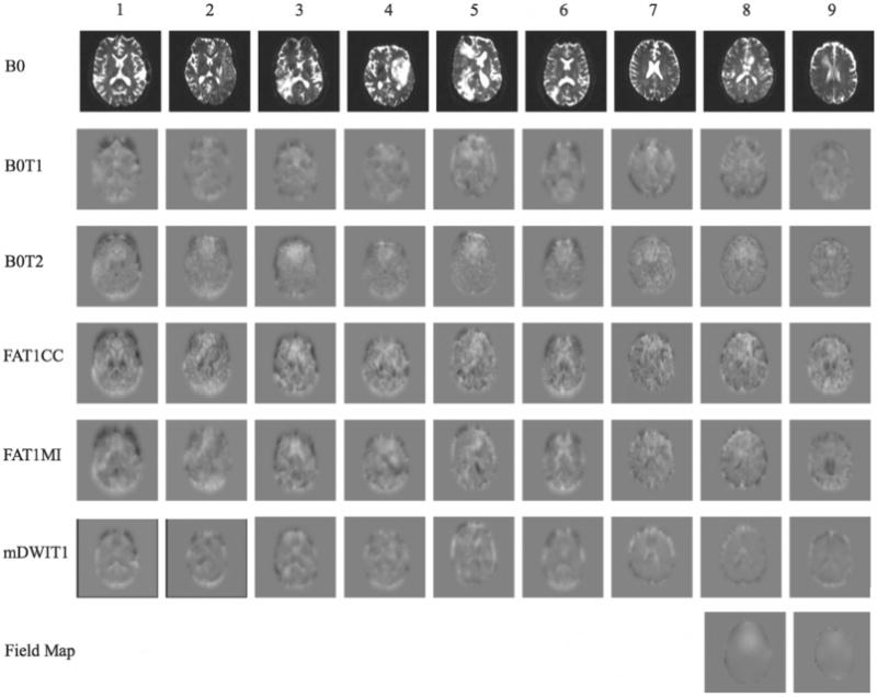
Methods: Five pipelines were tested in a retrospective clinical dMRI dataset of 9 patients with brain tumors. Pipelines differed in the choice of fixed and moving images and the similarity metric for image registration. Distortions were measured in two important tracts for neurosurgery, the arcuate fasciculus and corticospinal tracts.

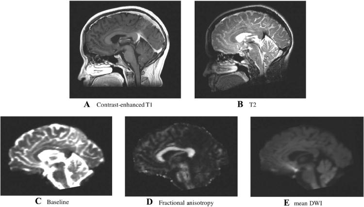
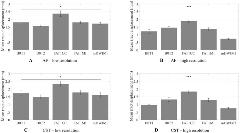
Results: Significant differences in distortion estimates were found across processing pipelines. The most successful pipeline used dMRI baseline and T2-weighted images as inputs for distortion correction. This pipeline gave the most consistent distortion estimates across image resolutions and brain hemispheres.

Conclusions: Quantitative results of mean tract distortions on the order of 1-2 mm are in line with other recent studies, supporting the potential need for distortion correction in neurosurgical planning. Novel results include significantly higher distortion estimates in the tumor hemisphere and greater effect of image resolution choice on results in the tumor hemisphere. Overall, this study demonstrates possible pitfalls and indicates that care should be taken when implementing EPI distortion correction in clinical settings.

Keywords: Diffusion tensor imaging; EPI distortion correction; image registration; neurosurgical planning; tractography.


Figures

Fig 1
Fig 1
Target and moving images. Anatomical MRI images used as registration targets included (A) T2-weighted and (B) contrast-enhanced T1-weighted. Images derived from diffusion MRI including (C) baseline, (D) fractional anisotropy, and (E) mean diffusion-weighted images were used as moving images in the EPI distortion correction experiments. Displayed images are from the first subject in the study. Note that a skull-stripping mask was applied to all images prior to deformable registration.
Fig 2
Fig 2
Mean absolute displacement measurement within anatomical tracts. Steps used to obtain measurements of the absolute displacement within the corticospinal tract (CST) and the arcuate fasciculus (AF, not shown). Each diffusion-weighted image (DWI) (A) underwent image distortion correction. From the distortion correction along the phase encoding direction, a deformation field (B) was obtained. The grayscale value indicates the amount of displacement with −15 mm in black and +15 mm in white. (C) Whole-brain tractography was performed on every distortion corrected volume and anatomical tracts such as CST (D) and AF were obtained. Within each of the tracts, we quantified the mean absolute displacement derived from the deformation field (E).
Fig 3
Fig 3
Estimated displacement in the phase encode direction. Estimated distortion using high-resolution data, where each column corresponds to one subject and each row corresponds to a pipeline. Deformation fields visualization is in the axial plane and indicates translation in the anterior-posterior direction (phase encode direction), as estimated by the registration pipelines. The grayscale value indicates the amount of displacement with −15 mm in black and +15 mm in white. Field maps: maps representing the field inhomogeneity across the images (available only for 2 subjects). B0 = baseline; B0T1 = baseline toT1- weighted anatomical image (T1) registration pipeline; B0T2 = baseline to T2-weighted anatomical image (T2) registration pipeline; FAT1CC = fractional anisotropy to T1 registration pipeline; FAT1MI = fractional anisotropy to T1 registration pipeline; mDWIT1 = mean diffusion-weighted image to T1 registration pipeline.
Fig 4
Fig 4
Spatial relationship of the tracts to the tumor. Spatial relationship of the tracts with the tumor: the distance from the tracts to the lesion can vary with the registration method. (A) 3D representation of tractography results in corticospinal tract from patient 9 with diffuse astrocytoma. After application of registration pipelines, the location of the seeded tracts appears visually similar in 3D. (B) Tracts from all pipelines, showing their intersection with a T2 sagittal slice. (C-I) Tracts’ intersection with a T2-weighted anatomical image shows the variability across pipelines. EC = Eddy current and movement corrected data; Uncorr = uncorrected data; B0T1 = baseline to T1-weighted anatomical image (T1) registration pipeline; B0T2 = baseline to T2-weighted anatomical image (T2) registration pipeline; FAT1CC = fractional anisotropy to T1 registration pipeline; FAT1MI = fractional anisotropy to T1 registration pipeline; mDWIT1 = mean diffusion-weighted image to T1 registration pipeline.
Fig 5
Fig 5
Estimated mean tract displacement. Comparison across registration pipelines shows significant differences in estimated displacement of critical white matter structures. Mean absolute displacement of arcuate fasciculus (low-resolution (A) and high-resolution (B) data) and corticospinal tract (low-resolution (C) and high-resolution (D) data), after averaging over hemisphere. Statistics were performed on mean displacement values (in mm) on N= 6 subjects for arcuate fasciculus (AF) and on N= 9 subjects for corticospinal tract (CST) using a one-way ANOVA (low-resolution data: AF, P= .012; CST, P= .014), * = P < .05; (high-resolution data: AF, P < .001; CST, P < .001), *** = P < .001. Significant comparisons are indicated by asterisks (*P < .05, **P < .001, ***P < .0001). B0T1 = baseline to T1-weighted anatomical image (T1) registration pipeline; B0T2 = baseline to T2-weighted anatomical image (T2) registration pipeline; FAT1CC = fractional anisotropy to T1 registration pipeline; FAT1MI = fractional anisotropy to T1 registration pipeline; mDWIT1 = mean diffusion-weighted image to T1 registration pipeline.
Fig 6
Fig 6
Effect of contrast enhancement on T1-based distortion correction method. Directionally encoded color images of the corpus callosum for one sample subject, showing the effect of contrast enhancement on the T1- based distortion correction method. (A) Fractional anisotropy map; (B-D) original contrast-enhanced T1-weighted image; (C) contrast-enhanced T1-weighted image with mask overlaid; (E) uncorrected DTI volume; (F) echo-planar imaging (EPI) distortion corrected data obtained with FAT1CC registration demonstrates “overstretching” of the corpus callosum; and (G) EPI distortion corrected data obtained with FAT1CC after masking brighter venous drainage resulting from the contrast enhancement in the T1-weighted volume.

References

    1. Jellison BJ, Field AS, Medow J, et al. Diffusion tensor imaging of cerebral white matter: a pictorial review of physics, fiber tract anatomy, and tumor imaging patterns. Am J Neuroradiol. 2005;25:356–69. - PMC - PubMed
    1. Wu JS, Zhou LF, Tang WJ, et al. Clinical evaluation and follow-up outcome of diffusion tensor imaging-based functional neuronavigation: a prospective, controlled study in patients with gliomas involving pyramidal tracts. Neurosurgery. 2007;61:935–49. - PubMed
    1. Jezzard P, Clare S. Sources of distortion in functional MRI data. Hum Brain Mapp. 1999;8:80–5. - PMC - PubMed
    1. Jezzard P, Balaban RS. Correction for geometric distortion in echo planar images from B0 field variations. Magn Reson Med. 1995;34:65–73. - PubMed
    1. Merhof D, Soza G, Stadlbauer A, et al. Correction of susceptibility artifacts in diffusion tensor data using non-linear registration. Med Image Anal. 2007;11:588–603. - PubMed

Publication types

LinkOut - more resources