. 2018 Apr 1;35(4):914-924.
doi: 10.1093/molbev/msy002.

Deuterostome Genomics: Lineage-Specific Protein Expansions That Enabled Chordate Muscle Evolution

Affiliations

Deuterostome Genomics: Lineage-Specific Protein Expansions That Enabled Chordate Muscle Evolution

Jun Inoue et al. Mol Biol Evol. .

Erratum in

Abstract

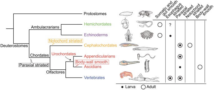
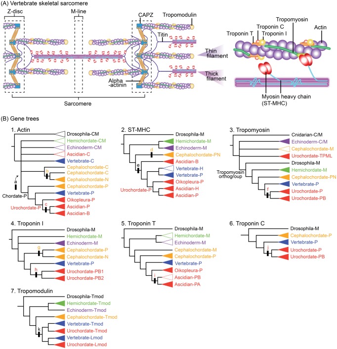
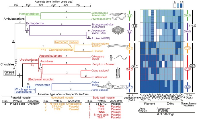
Fish-like larvae were foundational to the chordate body plan, given the basal placement of free-living lancelets. That body plan probably made it possible for chordate ancestors to swim by beating a tail formed of notochord and bilateral paraxial muscles. In order to investigate the molecular genetic basis of the origin and evolution of paraxial muscle, we deduced the evolutionary histories of 16 contractile protein genes from paraxial muscle, based on genomic data from all five deuterostome lineages, using a newly developed orthology identification pipeline and a species tree. As a result, we found that more than twice as many orthologs of paraxial muscle genes are present in chordates, as in nonchordate deuterostomes (ambulacrarians). Orthologs of paraxial-type actin and troponin C genes are absent in ambulacrarians and most paraxial muscle protein isoforms diversified via gene duplications that occurred in each chordate lineage. Analyses of genes with known expression sites indicated that some isoforms were reutilized in specific muscles of nonvertebrate chordates via gene duplications. As orthologs of most paraxial muscle genes were present in ambulacrarians, in addition to expression patterns of related genes and functions of the two protein isoforms, regulatory mechanisms of muscle genes should also be considered in future studies of the origin of paraxial muscle.


Figures

<sc>Fig</sc>. 1.
Fig. 1.
Phylogenetic relationships of major deuterostome lineages (Holland 1996; Simakov et al. 2015) and distribution of somatic and visceral muscle types (Schmidt-Rhaesa 2007). In hemichordates, most musculature exhibits smooth muscle structure (Benito and Pardos 1997) although the possible presence of esophagus striated muscles is suggested (Ceresa Castellani and Saita 1974). In echinoderms, histologically, muscle generally more closely resembles vertebrate smooth muscle than skeletal muscle (Garcia-Arraras and Dolmatov 2010). Striated structure is reported in brachial muscles of crinoids and the base of esophageal muscle of sea urchin larvae (Burke 1981). In cephalochordates, in addition to segmented myotomal musculature composed of striated muscle, notochord is filled with modified striated muscle cells (Holland 1996). In urochordates, appendicularians maintain the tail, consisting of a notochord, a nerve cord, and double bands of muscle cells throughout their lives (Nishino et al. 2000), whereas ascidians lose the larval tail and its muscle cells and form body-wall muscle in the trunk during metamorphosis into sessile adults (Burighel and Cloney 1997). Vertebrates possess skeletal and esophagus (Shiina et al. 2005) striated muscles, and visceral smooth muscles (Marieb and Hoehn 2015). Balloons in the species tree indicate phylogenetic positions of specific muscle origins inferred from the presence or absence of chordate muscle types (for discussion of visceral smooth muscle, see Oota and Saitou 1999; Brunet et al. 2016).
<sc>Fig</sc>. 2.
Fig. 2.
(A) Schematic representation of several molecules in vertebrate skeletal sarcomeres. Contraction occurs in response to calcium using troponin–tropomyosin regulatory mechanisms (Schmidt-Rhaesa 2007; Marieb and Hoehn 2015). (B) Schematic of estimated muscle protein gene trees (supplementary fig. S2AG, Supplementary Material online). Black bars with lower case letters indicate gene duplication events. Upper case letters in clade names indicate inferred expressions of ancestral genes (A, adult; B, body-wall muscle; C, cytoplasm; C/M, cytoplasm or muscle; H, heart; Lmod, leiomodin; M, muscle; N, notochord muscle; P, paraxial muscle; Tmod, tropomodulin; TPML, tropomyosin-like). Triangles indicate gene grouping in which monophyly is supported (closed) or not (open).
<sc>Fig</sc>. 3.
Fig. 3.
Time-calibrated deuterostome tree (Holland 1996; Simakov et al. 2015) and evolution of structural (contractile protein) genes of paraxial muscle as a summary of gene tree analyses (supplementary fig. S2AP, Supplementary Material online). Numbers at branches are counts of isoform families that experienced gene duplication and numbers at vertical bars (middle) are total counts of gene duplications in major lineages (supplementary fig. S3, Supplementary Material online). The heat map (right) reflects the estimated number of orthologs (table 1). Striped boxes in ambulacrarian troponin T indicate ambiguity of their functions as in chordate troponin T. The table (below) indicates inferred ancestral types of muscle-specific isoforms.

References

    1. Altschul SF, Madden TL, Schaffer AA, Zhang JH, Zhang Z, Miller W, Lipman DJ.. 1997. Gapped BLAST and PSI-BLAST: a new generation of protein database search programs. Nucleic Acids Res. 2517:3389–3402. - PMC - PubMed
    1. Andrikou C, Iovene E, Rizzo F, Oliveri P, Arnone MI.. 2013. Myogenesis in the sea urchin embryo: the molecular fingerprint of the myoblast precursors. Evodevo 41:33.. - PMC - PubMed
    1. Araki I, Saiga H, Makabe KW, Satoh N.. 1994. Expression of Amd1, a gene for a Myod1-related factor in the ascidian Halocynthia roretzi. Roux Arch Dev Biol. 2036:320–327. - PubMed
    1. Barnes DE, Hwang H, Ono K, Lu H, Ono S.. 2016. Molecular evolution of troponin I and a role of its N-terminal extension in nematode locomotion. Cytoskeleton (Hoboken) 733:117–130. 10.1002/cm.21281 - DOI - PMC - PubMed
    1. Benito J, Pardos F.. 1997. Hemichordata In: Harrison FW, Ruppert EE, editors. Microscopic anatomy of invertebrates. New York: Wiley-Liss Inc; p. 15–101.

Publication types

Substances