. 2018 Jan 10;15(1):104.
doi: 10.3390/ijerph15010104.

The Secretory Response of Rat Peritoneal Mast Cells on Exposure to Mineral Fibers

Affiliations

The Secretory Response of Rat Peritoneal Mast Cells on Exposure to Mineral Fibers

Violetta Borelli et al. Int J Environ Res Public Health. .

Abstract

Background: Exposure to mineral fibers is of substantial relevance to human health. A key event in exposure is the interaction with inflammatory cells and the subsequent generation of pro-inflammatory factors. Mast cells (MCs) have been shown to interact with titanium oxide (TiO₂) and asbestos fibers. In this study, we compared the response of rat peritoneal MCs challenged with the asbestos crocidolite and nanowires of TiO₂ to that induced by wollastonite employed as a control fiber.

Methods: Rat peritoneal MCs (RPMCs), isolated from peritoneal lavage, were incubated in the presence of mineral fibers. The quantities of secreted enzymes were evaluated together with the activity of fiber-associated enzymes. The ultrastructural morphology of fiber-interacting RPMCs was analyzed with electron microscopy.

Results: Asbestos and TiO₂ stimulate MC secretion. Secreted enzymes bind to fibers and exhibit higher activity. TiO₂ and wollastonite bind and improve enzyme activity, but to a lesser degree than crocidolite.

Conclusions: (1) Mineral fibers are able to stimulate the mast cell secretory process by both active (during membrane interaction) and/or passive (during membrane penetration) interaction; (2) fibers can be found to be associated with secreted enzymes-this process appears to create long-lasting pro-inflammatory environments and may represent the active contribution of MCs in maintaining the inflammatory process; (3) MCs and their enzymes should be considered as a therapeutic target in the pathogenesis of asbestos-induced lung inflammation; and (4) MCs can contribute to the inflammatory effect associated with selected engineered nanomaterials, such as TiO₂ nanoparticles.

Keywords: asbestos; crocidolite; endocytosis; mast cells; secretory process; titanium oxide nanowires.


Conflict of interest statement

The authors declare no conflict of interest. The founding sponsors had no role in the design of the study; in the collection, analyses, or interpretation of data; in the writing of the manuscript, and in the decision to publish the results.

Figures

Figure 1
Figure 1
Light microscope appearance of rat peritoneal MCs (RPMCs) exposed to mineral fibers or stimulated with compound 48/80. (a) Unstimulated RPMCs after 30 min of incubation; (b,c) RPMCs exposed to 100 µg/mL of crocidolite fibers after 5 or 30 min of incubation; (d) isolated RPMC membrane-covered granules incubated with 100 µg/mL of crocidolite for 30 min; (e) an RPMC exposed to 100 µg/mL TiO2 nanowires (NWs) after 30 min of incubation, arrowheads show intracellular nanowires and in the magnified inset the arrowhead shows a nanowire inside the cell; (f) RPMCs exposed to 100 µg/mL of wollastonite fibers after 30 min of incubation. ((g,h), inset) RPMCs stimulated with compound 48/80 10 µg/mL after 5 or 30 min incubation; the pictures are representative of at least ten different experiments. Cytocentrifuge-prepared samples were stained with the Diff-Quik system. Original magnification = 1000×.
Figure 1
Figure 1
Light microscope appearance of rat peritoneal MCs (RPMCs) exposed to mineral fibers or stimulated with compound 48/80. (a) Unstimulated RPMCs after 30 min of incubation; (b,c) RPMCs exposed to 100 µg/mL of crocidolite fibers after 5 or 30 min of incubation; (d) isolated RPMC membrane-covered granules incubated with 100 µg/mL of crocidolite for 30 min; (e) an RPMC exposed to 100 µg/mL TiO2 nanowires (NWs) after 30 min of incubation, arrowheads show intracellular nanowires and in the magnified inset the arrowhead shows a nanowire inside the cell; (f) RPMCs exposed to 100 µg/mL of wollastonite fibers after 30 min of incubation. ((g,h), inset) RPMCs stimulated with compound 48/80 10 µg/mL after 5 or 30 min incubation; the pictures are representative of at least ten different experiments. Cytocentrifuge-prepared samples were stained with the Diff-Quik system. Original magnification = 1000×.
Figure 2
Figure 2
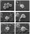
Scanning electron microscope appearance of RPMCs exposed to mineral fibers or stimulated with compound 48/80. (a) Unstimulated RPMCs after 30 min of incubation; (b) RPMCs exposed to 10 µg/mL compound 48/80 for 30 min: a large number of granules are secreted and the cell surface shows many protruding granules, while the inset shows an RPMC ghost induced by 48/80; (c,d) RPMC exposed to 100 µg/mL crocidolite for 30 min: an evident secretory response is shown, fiber-bound free granules as well as granule clusters can be seen, and numerous granule profiles protrude from the cell surface. ((d) and inset) Asbestos fibers can be seen, entrapped by the RPMC; the fibers may have reached the cell interior by either penetration or frustrated phagocytosis; (e) an RPMC exposed to 100 µg/mL TiO2NWs for 30 min. Note that numerous fibers interact with cell and free granules; ((f) and inset) an RPMC exposed to 100 µg/mL wollastonite fibers for 30 min. Note that despite many fibers being deposited on the RPMC surface there is no sign of degranulation. Magnification: bars in (a,b,e) = 3 µm; bars in (c) = 2 µm; in (d,f) = 1 µm.
Figure 3
Figure 3
Interaction of RPMC-isolated granules with crocidolite as observed by SEM. (a) Isolated membrane-covered granules alone; (b) isolated membrane-covered granules incubated with 100 µg/mL of crocidolite fibers; (c) isolated membrane-covered granules incubated with 100 µg/mL of TiO2NWs. A fiber-bound cluster of granules can be seen. Magnification: bars in (a) = 1 µm; (b) = 2 µm; (c) = 3 µm.
Figure 4
Figure 4
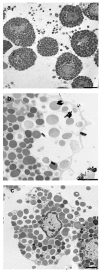
Transmission electron microscopy appearance of an RPMC exposed to 100 µg/mL of crocidolite fibers. After 30 min of incubation, the interacting fibers appear to undergo ingestion (a,b) or are free in the cytosol (arrowhead in (a,d)); (c) a degranulation process is evident where asbestos fibers are present; (d) the RPMC releasing a single granule (arrow). Magnifications: bar in (a) = 2 µm; bar in (b) = 1 µm; (c) = 2 µm; (d) = 0.5 µm.
Figure 5
Figure 5
Transmission electron microscopy appearance of RPMC exposed to 100 µg/mL of crocidolite fibers. (a) The unstimulated RPMC population; (b,c) a crocidolite-stimulated RPMC. After 30 min of incubation the secretory process appears to follow two pathways. In some cases, the secretion appears to follow the conventional compound exocytosis pathway (b), which is characterized by multiple granule fusion and formation of large vacuoles, which will subsequently release the granule content in one step and give rise to granule remnants. On bottom right a granule remnant adhering to asbestos fibers is shown. In other cells the process follows cell disruption with intact granule expulsion (c). Magnifications: bar in (a,c) = 2 µm.
Figure 6
Figure 6
Enzyme secretion from mineral fiber (100 µg/mL) or 48/80 compound (10 µg/mL)-stimulated RPMCs. Values are the mean ± SD of at least four different experiments. The extent of secretion, i.e., the percentage of enzyme activity in the supernatant, was calculated taking total enzyme activity (supernatant + pellet) as 100% (mean value ± standard deviation (SD): 0.074 ± 0.006 for chymase, 0.079 ± 0.006 for β-hexosaminidase (β-hexo) Optical Density (OD)/30 min and 0.060 ± 0.005 OD/30 min for tryptase). In the case of histamine, the total (supernatant + pellet) fluorescence arbitrary units were considered as 100%. See text for details. In the case of β-hexosaminidase, the control value did not differ significantly from that obtained with crocidolite stimulation. In all other cases, the difference between control and stimulated (crocidolite or 48/80) values was significantly different (p < 0.05) (asterisks). CTRL = control unstimulated cells; Cro = crocidolite-stimulated cells; Wolla = wollastonite-stimulated cells; TiO2 = TiO2NW-stimulated cells; 48/80 = 48/80-stimulated cells.
Figure 7
Figure 7
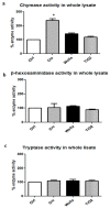
Effects of mineral fibers on enzyme activities in RPMC lysate. RPMC total lysate was obtained by sonication (3 × 10⁶ RPMC in 1 mL) and incubated for 30 min with mineral fibers (100 μg/mL). The values reported are the mean ± SD of at least five different experiments. The extent of enzyme activity was calculated taking the total enzyme activity present in the whole lysate as 100% (0.390 ± 0.052 OD/30 min for chymase; 0.160 ± 0.080 OD/30 min for β-hexosaminidase; 0120 ± 0.05 OD/30 min for tryptase (TRY)). Significant differences (p < 0.05) between unstimulated lysate and lysate from RPMC exposed to mineral fibers are indicated by asterisks. CTRL = unstimulated cells; Cro = crocidolite stimulated cells; Wolla = wollastonite-stimulated cells; TiO2 = TiO2NW-stimulated cells; 48/80 = 48/80-stimulated cells.
Figure 8
Figure 8
Extent of RPMC lysate enzyme absorption on mineral fibers. After 30 min incubation of the lysate in the presence of fibers (100 μg/mL), the supernatant was obtained by centrifuging the mixture at 250× g for 15 min at 4 °C. (a,b) fractionation of chymase activity; (c,d) fractionation of β-hexosaminidase activity. Significant changes in activity in the samples exposed to mineral fibers vs. baseline value (see the legend to Figure 7) are indicated by asterisks (p < 0.05). CTRL = control unstimulated cells; Cro = crocidolite-exposed enzyme; Wolla = wollastonite-exposed enzyme; TiO2 = TiO2NW-exposed enzyme.

Similar articles

Cited by

References

    1. Novello S., Pinto C., Torri V., Porcu L., Di Maio M., Tiseo M., Ceresoli G., Magnani C., Silvestri S., Veltri A., et al. The Third Italian Consensus Conference for Malignant Pleural Mesothelioma: State of the art and recommendations. Crit. Rev. Oncol. Hematol. 2016;104:9–20. doi: 10.1016/j.critrevonc.2016.05.004. - DOI - PubMed
    1. Ferin J., Oberdörster G., Penney D.P. Pulmonary retention of ultrafine and fine particles in rats. Am. J. Respir. Cell Mol. Biol. 1992;6:535–542. doi: 10.1165/ajrcmb/6.5.535. - DOI - PubMed
    1. Magrez A., Horváth L., Smajda R., Salicio V., Pasquier N., Forró L., Schwaller B. Cellular toxicity of TiO2-based nanofilaments. ACS Nano. 2009;3:2274–2280. doi: 10.1021/nn9002067. - DOI - PubMed
    1. Park E.J., Shim H.W., Lee G.H., Kim J.H., Kim D.W. Comparison of toxicity between the different-type TiO2 nanowires in vivo and in vitro. Arch. Toxicol. 2013;87:1219–1230. doi: 10.1007/s00204-013-1019-3. - DOI - PubMed
    1. Wang J., Fan Y. Lung Injury Induced by TiO2 Nanoparticles Depends on Their Structural Features: Size, Shape, Crystal Phases and Surface Coating. Int. J. Mol. Sci. 2014;15:22258–22278. doi: 10.3390/ijms151222258. - DOI - PMC - PubMed

Publication types

LinkOut - more resources