

Functional Testing of SLC26A4 Variants-Clinical and Molecular Analysis of a Cohort with Enlarged Vestibular Aqueduct from Austria
- PMID: 29320412
- PMCID: PMC5796158
- DOI: 10.3390/ijms19010209
Functional Testing of SLC26A4 Variants-Clinical and Molecular Analysis of a Cohort with Enlarged Vestibular Aqueduct from Austria
Abstract
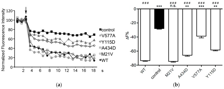
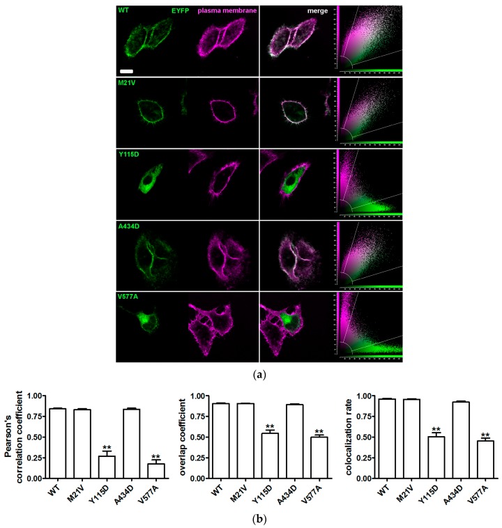
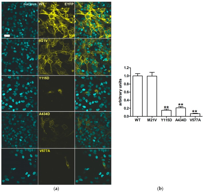
The prevalence and spectrum of sequence alterations in the SLC26A4 gene, which codes for the anion exchanger pendrin, are population-specific and account for at least 50% of cases of non-syndromic hearing loss associated with an enlarged vestibular aqueduct. A cohort of nineteen patients from Austria with hearing loss and a radiological alteration of the vestibular aqueduct underwent Sanger sequencing of SLC26A4 and GJB2, coding for connexin 26. The pathogenicity of sequence alterations detected was assessed by determining ion transport and molecular features of the corresponding SLC26A4 protein variants. In this group, four uncharacterized sequence alterations within the SLC26A4 coding region were found. Three of these lead to protein variants with abnormal functional and molecular features, while one should be considered with no pathogenic potential. Pathogenic SLC26A4 sequence alterations were only found in 12% of patients. SLC26A4 sequence alterations commonly found in other Caucasian populations were not detected. This survey represents the first study on the prevalence and spectrum of SLC26A4 sequence alterations in an Austrian cohort and further suggests that genetic testing should always be integrated with functional characterization and determination of the molecular features of protein variants in order to unequivocally identify or exclude a causal link between genotype and phenotype.
Keywords: SLC26A4; enlarged vestibular aqueduct; non-syndromic hearing loss.
Conflict of interest statement
The authors declare no conflict of interest.
Figures


Similar articles
-
Novel genetic determinants contribute to hearing loss in a central European cohort with enlarged vestibular aqueduct.Mol Med. 2025 Mar 22;31(1):111. doi: 10.1186/s10020-025-01159-9. Mol Med. 2025. PMID: 40121402 Free PMC article.
-
Genetic heterogeneity in patients with enlarged vestibular aqueduct and Pendred syndrome.Mol Med. 2025 May 27;31(1):208. doi: 10.1186/s10020-025-01262-x. Mol Med. 2025. PMID: 40426046 Free PMC article.
-
SLC26A4 gene is frequently involved in nonsyndromic hearing impairment with enlarged vestibular aqueduct in Caucasian populations.Eur J Hum Genet. 2006 Jun;14(6):773-9. doi: 10.1038/sj.ejhg.5201611. Eur J Hum Genet. 2006. PMID: 16570074
-
Molecular and functional characterization of human pendrin and its allelic variants.Cell Physiol Biochem. 2011;28(3):451-66. doi: 10.1159/000335107. Epub 2011 Nov 18. Cell Physiol Biochem. 2011. PMID: 22116358 Review.
-
Genetic architecture and phenotypic landscape of SLC26A4-related hearing loss.Hum Genet. 2022 Apr;141(3-4):455-464. doi: 10.1007/s00439-021-02311-1. Epub 2021 Aug 3. Hum Genet. 2022. PMID: 34345941 Review.
Cited by
-
Genetics of Inner Ear Malformations: A Review.Audiol Res. 2021 Oct 12;11(4):524-536. doi: 10.3390/audiolres11040047. Audiol Res. 2021. PMID: 34698066 Free PMC article. Review.
-
Novel genetic determinants contribute to hearing loss in a central European cohort with enlarged vestibular aqueduct.Mol Med. 2025 Mar 22;31(1):111. doi: 10.1186/s10020-025-01159-9. Mol Med. 2025. PMID: 40121402 Free PMC article.
-
Clinical and Molecular Aspects Associated with Defects in the Transcription Factor POU3F4: A Review.Biomedicines. 2023 Jun 12;11(6):1695. doi: 10.3390/biomedicines11061695. Biomedicines. 2023. PMID: 37371790 Free PMC article. Review.
-
Inhibitors of the ubiquitin‑proteasome system rescue cellular levels and ion transport function of pathogenic pendrin (SLC26A4) protein variants.Int J Mol Med. 2025 May;55(5):69. doi: 10.3892/ijmm.2025.5510. Epub 2025 Mar 7. Int J Mol Med. 2025. PMID: 40052591 Free PMC article.
-
Molecular Features of SLC26A4 Common Variant p.L117F.J Clin Med. 2022 Sep 22;11(19):5549. doi: 10.3390/jcm11195549. J Clin Med. 2022. PMID: 36233414 Free PMC article.
References
-
- Da Silva Costa S.M., Ramos P.Z., Arrojo Martins F.T., Sartorato E.L. Genetic Diagnosis of Deafness. In: Dossena S., Paulmichl M., editors. The Role of Pendrin in Health and Disease. Springer International Publishing; Basel, Switzerland: 2017. pp. 61–83.
-
- Alasti F., Van Camp G., Smith R.J.H. Pendred Syndrome/DFNB4. In: Pagon R.A., Adam M.P., Ardinger H.H., Wallace S.E., Amemiya A., Bean L.J.H., Bird T.D., Ledbetter N., Mefford H.C., Smith R.J.H., et al., editors. GeneReviews(R) University of Washington; Seattle, WA, USA: 1993.
MeSH terms
Substances
Supplementary concepts
LinkOut - more resources
Full Text Sources
Other Literature Sources
