. 2018 Jan 18;553(7688):361-365.
doi: 10.1038/nature25190. Epub 2018 Jan 10.

Architecture of a channel-forming O-antigen polysaccharide ABC transporter

Affiliations

Architecture of a channel-forming O-antigen polysaccharide ABC transporter

Yunchen Bi et al. Nature. .

Abstract

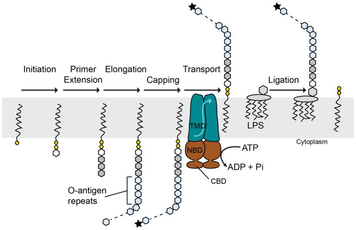
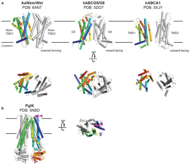
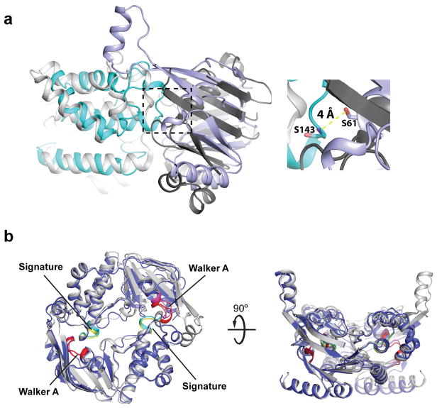
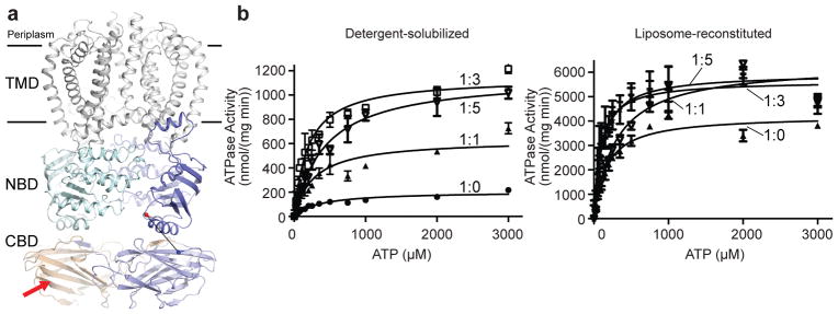
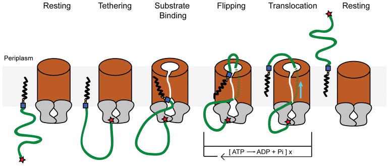
O-antigens are cell surface polysaccharides of many Gram-negative pathogens that aid in escaping innate immune responses. A widespread O-antigen biosynthesis mechanism involves the synthesis of the lipid-anchored polymer on the cytosolic face of the inner membrane, followed by transport to the periplasmic side where it is ligated to the lipid A core to complete a lipopolysaccharide molecule. In this pathway, transport to the periplasm is mediated by an ATP-binding cassette (ABC) transporter, called Wzm-Wzt. Here we present the crystal structure of the Wzm-Wzt homologue from Aquifex aeolicus in an open conformation. The transporter forms a transmembrane channel that is sufficiently wide to accommodate a linear polysaccharide. Its nucleotide-binding domain and a periplasmic extension form 'gate helices' at the cytosolic and periplasmic membrane interfaces that probably serve as substrate entry and exit points. Site-directed mutagenesis of the gates impairs in vivo O-antigen secretion in the Escherichia coli prototype. Combined with a closed structure of the isolated nucleotide-binding domains, our structural and functional analyses suggest a processive O-antigen translocation mechanism, which stands in contrast to the classical alternating access mechanism of ABC transporters.


Conflict of interest statement

Competing financial interests

The authors declare no competing financial interests

Figures

Extended Data Figure 1
Extended Data Figure 1. ABC transporter-dependent O-antigen biosynthesis
In this pathway, O antigens are completely synthesized on the cytosolic leaflet of the plasma membrane. Undecaprenyl-phosphate (black line and yellow circle) serves as the lipid acceptor and is modified by the addition of an acetylated amino sugar phosphate (frequently N-acetylglucosamine-1-P, white hexagon) as well as 2 or more additional sugar residues (gray hexagons) to generate a biosynthesis primer. The polymerizing enzyme(s) extend the primer with tens to hundreds of O-antigen repeat units (light blue hexagons). In some species, termination of O antigen biosynthesis is achieved by modifying the polymer’s non-reducing end (black star). An ABC transporter translocates the Und-PP-linked O antigen intermediate to the membrane’s periplasmic side, where it forms a substrate for glycosylation of the lipopolysaccharide (LPS) core. Only transporters translocating terminally-modified O-antigens contain carbohydrate-binding domains (CBD) that bind the polysaccharide’s modified terminus. TMD/NBD: Transmembrane and nucleotide binding domains.
Extended Data Figure 2
Extended Data Figure 2. Sequence alignment of O antigen and wall teichoic acid transporters
Alignments of the nucleotide binding and transmembrane domains are shown in panels (a) and (b), respectively. The conserved tyrosine preceding the NBD’s cytosolic gate helix and in the periplasmic gate are highlighted with a red arrow and red box in panels a and b, respectively. Transmembrane helices and cytosolic and periplasmic gate helices are shown as green and beige cylinders, respectively. Blue sequence labels indicate predicted teichoic acid transporters. All O antigen transporter NBDs except for K. pneumoniae O2a contain predicted CBDs at their C termini, which are not shown. (c) Alignment of the C-terminal region of AaWzt with the corresponding domains from the E. coli O9a (PDB: 2R5O) and R. terrigena/K. pneumoniae O12 (PDB: 5HNO) transporters. Sequences were aligned in CLUSTAL Omega and displayed in Jalview colored by sequence identity.
Extended Data Figure 3
Extended Data Figure 3. Anomalous difference and experimental electron density maps
(a) Heavy atom positions used for experimental phasing and model building. Five native cysteines in the NBDs as well as an engineered Cys at the C terminus of TM3 (T128C) were modified with ethylmercurithiosialicylic acid, shown as green and red meshes and contoured at 4.5 and 3σ, respectively. Only the Hg sites shown in green were used for Hg-SAD phasing. The TMD contains three native Met residues, which were identified upon substitution with seleno-methionine (cyan mesh, contoured at 3σ). Shown are SigmaA-weighted anomalous difference electron densities, AaWzm/WztN is shown as a gray ribbon. (b) Unbiased experimental SigmaA-weighted electron density after NCS and cross-crystal averaging and phase extension to 3.85Å, contoured at 1σ.
Extended Data Figure 4
Extended Data Figure 4. Comparison of Type-II ABC exporter folds
(a) The structures of the transmembrane domains of A. aeolicus Wzm, H. sapiens ABCG5 and H. sapiens ABCA1 are shown as cylindrical cartoons. One subunit of the dimers is colored in rainbow colors from blue to red, N to C terminus. (b) Structure of PglK, an ABC transporter translocating Und-PP-linked oligosaccharides across the plasma membrane. PglK likely recognizes the substrate’s polyprenyl moiety via a conserved periplasmic helix (shown in magenta), which is missing in Wzm.
Extended Data Figure 5
Extended Data Figure 5. Closed conformation of the isolated WztN NBD
(a) The isolated WztN dimer structure was aligned by secondary matching in Coot with the NBDs of the AMPPNP-stabilized maltose transporter, PDB entry 3RLF. The WztN dimer is shown in cyan and light blue and the maltose transporter’s NBDs are shown in light and dark gray. Right panel: The Walker A (S61) and Signature (S143) motifs in the closed WztN dimer structure are separated by approx. 4 Å. (b) Comparison of WztN dimer structures. The structure shown in dark blue was obtained from a crystal form containing a WztN dimer in the crystallographic asymmetric unit. The structure shown in gray was obtained from a crystal form with a monomeric WztN per crystallographic asymmetric unit related to the other protomer by two-fold crystallographic symmetry. The Signature motifs are colored cyan and yellow and the Walker-A motifs are colored magenta and red for the crystallographic monomeric and dimeric WztN structures, respectively.
Extended Data Figure 6
Extended Data Figure 6. Impact of conserved tyrosine residues of the cytosolic and periplasmic gates on O antigen translocation
The indicated point mutations were introduced into the E. coli O9a Wzt/Wzm transporter and O antigen transport was assayed by silver staining of the whole-cell lysate. Ag: Silver stained SDS-PAGE. Wzt and MBP were detected immunologically to monitor transporter expression and as a loading control, respectively. All experiments were repeated independently at least three times with similar results. Time: Period after inducing Wzt/Wzm expression in minutes.
Extended Data Figure 7
Extended Data Figure 7. Dimerization of the isolated Wzt carbohydrate-binding domain
Multi-angle static light scattering coupled to size-exclusion chromatography was used to determine the molecular weight of the purified Wzt carbohydrate-binding domain (one representative experiment is shown). The molecular weight of a monomeric Wzt-CBD is 20 kDa, including a C-terminal 6x-His-tag and linker region. Inset: Coomassie-stained SDS-PAGE of purified Wzt-CBD.
Extended Data Figure 8
Extended Data Figure 8. Hydrolytic activity of the Wzm/Wzt ABC transporter
ATP hydrolytic activity was measured by following the decrease of NADH fluorescence in an enzyme-coupled assay upon excitation at 340 nm and emission at 450 nm in a temperature range from 4 to 65°C. (a) Temperature dependence of Wzm/WztN’s ATPase activity. Shown is the difference in NADH fluorescence between control reactions in the absence of Wzm/WztN and reactions in its presence. (b) Hydrolytic activity of full-length Wzm/Wzt in the presence of isolated Wzt-CBD measured at 27°C. Shown are fluorescence intensity differences (calculated as for Fig. 4b) but not converted to apparent catalytic rates. Dashed line: ATP titration in the presence of only the Wzt-CBD. Hydrolytic activity of Wzm/WztN in the absence of Wzt-CBD is shown for comparison. (c) Comparison of ATPase activities of full-length (green) and truncated (black) Wzm/Wzt. Shown are apparent catalytic rates in detergent-solubilized and liposome-reconstitute states. Data points represent the mean of a three independent repeats with standard deviations. CPS: Counts per second.
Extended Data Figure 9
Extended Data Figure 9. Model of the Wzm/WztN closed conformation
(a) Rigid body alignment of the Wzm/WztN transporter halves with the corresponding NBDs of the closed WztN dimer structure. The closed WztN dimer is shown in gray and Wzm/WztN is colored in red and green for Wzm and cyan and blue for WztN. Residues replaced with Cys are shown as spheres for their C-alpha carbons and labeled. Observed disulfide cross-links are indicated with a dashed line. (b) Cartoon illustration of the transporter’s open to closed transition. (c) Disulfide cross-linking of Wzm protomers. Purified Wzm/WztN transporters harboring the indicated Cys mutations were oxidized either with copper-phenanthroline (Co-Phen) or sodium tetrathionate (STT), blocked with N-ethylmaleimide (NEM), and analyzed by Western blotting against the N-terminal Wzm FLAG-tag. Experiments were repeated three times with similar results. M and D: Wzm monomer and dimer.
Figure 1
Figure 1. Architecture of the Wzm/Wzt O antigen transporter
(a) The Wzm protomers are shown in green and red and the nucleotide-binding WztN domains are shown in blue and gray, respectively. WztN forms a short gate helix (GH) near the Wzm protomer interface. Wzm contains an N-terminal interface helix (IF). (b) Transmembrane topology of Wzm. Wzm forms six transmembrane helices and the cytosolic TM2/3 loop forms the coupling helix (CH) Wzm’s periplasmic TM5/6 loop generates two periplasmic gate helices (PG1 and 2). Horizontal lines indicate likely membrane boundaries.
Figure 2
Figure 2. Wzt forms a unique interface with Wzm
(a) Position of the gate helix (GH) at the Wzm protomer interface. The GH packs against Wzm’s TM4/5 loop and forms a wedge-shaped opening towards the cytosolic water-lipid interface. The transporter is shown as a cartoon and one Wzm protomer is shown as a semi-transparent surface. (b) Open conformation of the WztN NBDs. Surface representation of the transporter’s NBDs colored blue and cyan, respectively. Conserved regions are labeled. (c) Surface representation of the isolated WztN structure in a closed conformation colored as in (b).
Figure 3
Figure 3. The polysaccharide translocation channel
(a) The Wzm interface. One Wzm protomer is shown as a surface and the opposing subunit as a cartoon. Both subunits are shown as cartoons in the close-up view. TM1 and TM5 are colored red and green, respectively, and the IF is colored beige. Conserved residues are shown as sticks. (b) Surface representation of the Wzm/WztN channel. The channel volume accessible to a 3.5 Å radius probe is shown as a green surface and aromatic residues lining the channel are shown as brown spheres. Selected residues are labeled. (c) Cytosolic and periplasmic gate helices at the Wzm protomer interface. A model of the E. coli O9a antigen containing 10 mannose units was manually placed in the channel and is shown as a red surface. (d) Putative translocation path (red dashed line). Channel-exposed aromatic and polar residues are shown as brown sticks. (e) In vivo O antigen translocation. The indicated point mutations were introduced into E. coli O9a Wzm/Wzt. O antigen export was detected after inducing transporter expression by silver staining (Ag) of whole-cell lysates, detecting exported and LPS-linked O antigens only. Western blots detecting Wzt and MBP were performed to monitor transporter expression and as a loading control, respectively. All experiments were repeated independently at least three times with similar results. Time=Period after inducing Wzt/Wzm expression.
Figure 4
Figure 4. The CBD stimulates Wzm/WztN’s hydrolytic activity
(a) Putative organization of the full-length Wzm/Wzt transporter. Alignment of the E. coli O9a Wzt CBD structure (PDB: 2R5O) with the Wzm/WztN transporter. C and N termini of WztN and CBD are shown as red and blue spheres, respectively. Red arrow: putative binding site of the modified O antigen cap. (b) Hydrolytic activity of Wzm/WztN in detergent-solubilized and lipid-reconstituted states, respectively. ATP hydrolysis was performed under increasing CBD concentrations as indicated (Wzm/WztN:CBD, molar ratio). Error bars represent the standard deviations from the means of at least three independent replicas.
Figure 5
Figure 5. Model of O antigen membrane translocation
In a resting state, the transporter’s TM channel and the NBDs are in a closed conformation. Tethering the substrate to the transporter via interactions of the CBD with the modified O antigen terminus increases its local concentration. Binding of the Und-PP lipid anchor to the cytosolic gate induces NBD and TM channel opening. The lipid head group inserts into the channel and reorients spontaneously to the periplasmic side. The now channel-inserted polysaccharide is translocated through repeated cycles of ATP binding and hydrolysis. Upon polymer release to the periplasmic side, the transporter returns to the resting conformation with a closed TM channel. Blue square: N-acetylglucosamine; yellow spheres: phosphate; red star: modified terminus.

References

    1. Whitfield C, Szymanski CM, Aebi M. In: Essentials of Glycobiology. Varki A, editor. Chapter 21. 2017. pp. 265–282.
    1. Raetz CRH, Whitfield C. Lipopolysaccharide endotoxins. Annu Rev Biochem. 2002;71:635–700. - PMC - PubMed
    1. Caboni M, et al. An O antigen capsule modulates bacterial pathogenesis in Shigella sonnei. PLoS Pathog. 2015;11:e1004749. - PMC - PubMed
    1. Goebel EM, Wolfe DN, Elder K, Stibitz S, Harvill ET. O antigen protects Bordetella parapertussis from complement. Infect Immun. 2008;76:1774–1780. - PMC - PubMed
    1. Skurnik M, Bengoechea JA. The biosynthesis and biological role of lipopolysaccharide O-antigens of pathogenic Yersiniae. Carbohydr Res. 2003;338:2521–2529. - PubMed

Publication types

MeSH terms