

Pattern of peripapillary capillary density loss in ischemic optic neuropathy compared to that in primary open-angle glaucoma
- PMID: 29320503
- PMCID: PMC5761857
- DOI: 10.1371/journal.pone.0189237
Pattern of peripapillary capillary density loss in ischemic optic neuropathy compared to that in primary open-angle glaucoma
Abstract
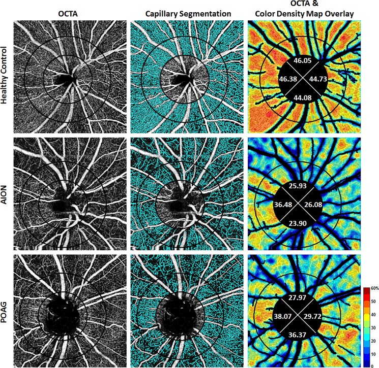
Purpose: Both non-arteritic anterior ischemic optic neuropathy (NAION) and primary open-angle glaucoma (POAG) damage retinal ganglion cell axons, which are perfused by the radial peripapillary capillaries. To evaluate the pattern of ischemia, we compared peripapillary capillary density (PCD) in NAION eyes to POAG eyes matched for visual field mean deviation and retinal nerve fiber layer thickness.
Methods: 31 chronic NAION (>6 months after the acute event) and unaffected fellow eyes (31 subjects), 42 moderate and severe POAG eyes (27 subjects), and 77 control eyes (46 healthy subjects) were imaged with a commercial optical coherence tomography angiography system (AngioVue, Avanti RTVue-XR, Optovue, CA) at two academic institutions. Two concentric circles of diameters 1.95mm (inner) and 3.45mm (outer) were manually placed on images centered on the optic nerve head, producing an annular region-of-interest. Image analysis with major vessel removal was performed using a custom program. Whole-image, whole-annulus, and sectoral PCDs were measured.
Results: Whole-image and whole-annulus PCDs in NAION and moderate and severe POAG eyes were significantly decreased compared to unaffected fellow eyes and control eyes (all P<0.001). Superior and temporal PCD values were affected more than other sectors in both NAION and POAG groups compared to control group. Whole-image and whole-annulus PCDs were not statistically different between NAION and POAG eyes (both P = 0.99). However, of all peripapillary sectors, the inferior sector PCD value was less affected in POAG eyes compared to NAION eyes (P = 0.001). Univariate analysis results also revealed a significant positive correlation between superior and inferior PCDs and corresponding RNFL thicknesses. The inferior sector correlation was greater in POAG than NAION eyes.
Conclusion: While the whole PCD values were not different in chronic NAION and POAG, the greater correlation of inferior PCD with corresponding RNFL sectors in POAG compared to NAION suggests greater susceptibility of the inferior radial peripapillary capillary in the pathogenesis of POAG.
Conflict of interest statement
Figures
Similar articles
-
Comparison of the Pattern of Macular Ganglion Cell-Inner Plexiform Layer Defect Between Ischemic Optic Neuropathy and Open-Angle Glaucoma.Invest Ophthalmol Vis Sci. 2016 Mar;57(3):1011-6. doi: 10.1167/iovs.15-18618. Invest Ophthalmol Vis Sci. 2016. PMID: 26962697
-
Microvascular and structural alterations in the optic nerve head of advanced primary open-angle glaucoma compared with atrophic non-arteritic anterior ischemic optic neuropathy.Graefes Arch Clin Exp Ophthalmol. 2021 Jul;259(7):1945-1953. doi: 10.1007/s00417-021-05122-2. Epub 2021 Mar 4. Graefes Arch Clin Exp Ophthalmol. 2021. PMID: 33661365
-
Peripapillary Perfused Capillary Density in Exfoliation Syndrome and Exfoliation Glaucoma versus POAG and Healthy Controls: An OCTA Study.Asia Pac J Ophthalmol (Phila). 2018 Mar-Apr;7(2):84-89. doi: 10.22608/APO.2017318. Epub 2017 Nov 22. Asia Pac J Ophthalmol (Phila). 2018. PMID: 29165935
-
Microvascular alterations detected by optical coherence tomography angiography in non-arteritic anterior ischaemic optic neuropathy: a meta-analysis.Acta Ophthalmol. 2022 Mar;100(2):e386-e395. doi: 10.1111/aos.14930. Epub 2021 Jun 21. Acta Ophthalmol. 2022. PMID: 34155823
-
[Nonarteritic ischemic optic neuropathy animal model and its treatment applications].Nippon Ganka Gakkai Zasshi. 2014 Apr;118(4):331-61. Nippon Ganka Gakkai Zasshi. 2014. PMID: 24864434 Review. Japanese.
Cited by
-
Effect of Smoking on Retinal Thickness and Vascular Density in Thyroid Eye Disease.Korean J Ophthalmol. 2021 Oct;35(5):376-382. doi: 10.3341/kjo.2021.0059. Epub 2021 Aug 3. Korean J Ophthalmol. 2021. PMID: 34344134 Free PMC article.
-
A Pilot Study of Subclinical Non-Capillary Peripapillary Perfusion Changes in Thyroid-Related Orbitopathy Detected Using Optical Coherence Tomography Angiography.Clin Ophthalmol. 2022 Mar 20;16:867-875. doi: 10.2147/OPTH.S356631. eCollection 2022. Clin Ophthalmol. 2022. PMID: 35340669 Free PMC article.
-
Automated Evaluation of Parapapillary Choroidal Microvasculature in Ischemic Optic Neuropathy and Open Angle Glaucoma.Invest Ophthalmol Vis Sci. 2020 Mar 9;61(3):35. doi: 10.1167/iovs.61.3.35. Invest Ophthalmol Vis Sci. 2020. PMID: 32191289 Free PMC article.
-
Early Macular Vessel Density Loss in Acute Ischemic Optic Neuropathy Compared to Papilledema: Implications for Pathogenesis.Transl Vis Sci Technol. 2018 Sep 26;7(5):10. doi: 10.1167/tvst.7.5.10. eCollection 2018 Sep. Transl Vis Sci Technol. 2018. PMID: 30271677 Free PMC article.
-
Comparison of Optic Nerve Head Microvasculature Between Normal-Tension Glaucoma and Nonarteritic Anterior Ischemic Optic Neuropathy.Invest Ophthalmol Vis Sci. 2021 Aug 2;62(10):15. doi: 10.1167/iovs.62.10.15. Invest Ophthalmol Vis Sci. 2021. PMID: 34398197 Free PMC article.
References
-
- Arnold AC. Ischemic optic neuropathy In: Miller NR, Newman NJ, Biousse V, editors. Clinical Neuro-Ophthalmology. 6th ed Vol. 1 Philadelphia, PA: Lippincott Williams & Wilkins; 2005. pp 349–84.
-
- Fard MA, Afzali M, Abdi P, Yasseri M, Ebrahimi KB, Moghimi S. Comparison of the pattern of macular ganglion cell-inner plexiform layer defect between ischemic optic neuropathy and open–angle glaucoma. Invest Ophthalmol Vis Sci 2016;57:1011–1016. doi: 10.1167/iovs.15-18618 - DOI - PubMed
-
- Petrig BL, Riva CE, Hayreh SS. Laser Doppler flowmetry and optic nerve head blood flow. Am J Ophthalmol 1999;127:413–425. - PubMed
-
- Sugiyama T, Araie M, Riva CE, Schmetterer L, Orgul S. Use of laser speckle flowgraphy in ocular blood flow research. Acta Ophthalmol (Copenh.) 2010; 88: 723–729. - PubMed
-
- Jia Y, Wei E, Wang X, Zhang X, Morrison JC, Parikh M, et al. Optical coherence tomography angiography of optic disc perfusion in glaucoma. Ophthalmology 2014;121:1322–1332. doi: 10.1016/j.ophtha.2014.01.021 - DOI - PMC - PubMed
Publication types
MeSH terms
LinkOut - more resources
Full Text Sources
Other Literature Sources
