

Pattern of peripapillary capillary density loss in ischemic optic neuropathy compared to that in primary open-angle glaucoma
- PMID: 29320503
- PMCID: PMC5761857
- DOI: 10.1371/journal.pone.0189237
Pattern of peripapillary capillary density loss in ischemic optic neuropathy compared to that in primary open-angle glaucoma
Abstract
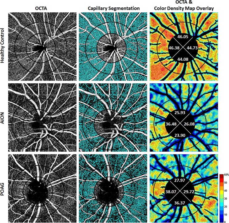
Purpose: Both non-arteritic anterior ischemic optic neuropathy (NAION) and primary open-angle glaucoma (POAG) damage retinal ganglion cell axons, which are perfused by the radial peripapillary capillaries. To evaluate the pattern of ischemia, we compared peripapillary capillary density (PCD) in NAION eyes to POAG eyes matched for visual field mean deviation and retinal nerve fiber layer thickness.
Methods: 31 chronic NAION (>6 months after the acute event) and unaffected fellow eyes (31 subjects), 42 moderate and severe POAG eyes (27 subjects), and 77 control eyes (46 healthy subjects) were imaged with a commercial optical coherence tomography angiography system (AngioVue, Avanti RTVue-XR, Optovue, CA) at two academic institutions. Two concentric circles of diameters 1.95mm (inner) and 3.45mm (outer) were manually placed on images centered on the optic nerve head, producing an annular region-of-interest. Image analysis with major vessel removal was performed using a custom program. Whole-image, whole-annulus, and sectoral PCDs were measured.
Results: Whole-image and whole-annulus PCDs in NAION and moderate and severe POAG eyes were significantly decreased compared to unaffected fellow eyes and control eyes (all P<0.001). Superior and temporal PCD values were affected more than other sectors in both NAION and POAG groups compared to control group. Whole-image and whole-annulus PCDs were not statistically different between NAION and POAG eyes (both P = 0.99). However, of all peripapillary sectors, the inferior sector PCD value was less affected in POAG eyes compared to NAION eyes (P = 0.001). Univariate analysis results also revealed a significant positive correlation between superior and inferior PCDs and corresponding RNFL thicknesses. The inferior sector correlation was greater in POAG than NAION eyes.
Conclusion: While the whole PCD values were not different in chronic NAION and POAG, the greater correlation of inferior PCD with corresponding RNFL sectors in POAG compared to NAION suggests greater susceptibility of the inferior radial peripapillary capillary in the pathogenesis of POAG.
Conflict of interest statement
Figures
References
-
- Arnold AC. Ischemic optic neuropathy In: Miller NR, Newman NJ, Biousse V, editors. Clinical Neuro-Ophthalmology. 6th ed Vol. 1 Philadelphia, PA: Lippincott Williams & Wilkins; 2005. pp 349–84.
-
- Fard MA, Afzali M, Abdi P, Yasseri M, Ebrahimi KB, Moghimi S. Comparison of the pattern of macular ganglion cell-inner plexiform layer defect between ischemic optic neuropathy and open–angle glaucoma. Invest Ophthalmol Vis Sci 2016;57:1011–1016. doi: 10.1167/iovs.15-18618 - DOI - PubMed
-
- Petrig BL, Riva CE, Hayreh SS. Laser Doppler flowmetry and optic nerve head blood flow. Am J Ophthalmol 1999;127:413–425. - PubMed
-
- Sugiyama T, Araie M, Riva CE, Schmetterer L, Orgul S. Use of laser speckle flowgraphy in ocular blood flow research. Acta Ophthalmol (Copenh.) 2010; 88: 723–729. - PubMed
-
- Jia Y, Wei E, Wang X, Zhang X, Morrison JC, Parikh M, et al. Optical coherence tomography angiography of optic disc perfusion in glaucoma. Ophthalmology 2014;121:1322–1332. doi: 10.1016/j.ophtha.2014.01.021 - DOI - PMC - PubMed
Publication types
MeSH terms
LinkOut - more resources
Full Text Sources
Other Literature Sources
