. 2018 Jan 10;13(1):e0189985.
doi: 10.1371/journal.pone.0189985. eCollection 2018.

Developmental finite element analysis of cichlid pharyngeal jaws: Quantifying the generation of a key innovation

Affiliations

Developmental finite element analysis of cichlid pharyngeal jaws: Quantifying the generation of a key innovation

Tim Peterson et al. PLoS One. .

Erratum in

Abstract

Advances in imaging and modeling facilitate the calculation of biomechanical forces in biological specimens. These factors play a significant role during ontogenetic development of cichlid pharyngeal jaws, a key innovation responsible for one of the most prolific species diversifications in recent times. MicroCT imaging of radiopaque-stained vertebrate embryos were used to accurately capture the spatial relationships of the pharyngeal jaw apparatus in two cichlid species (Haplochromis elegans and Amatitlania nigrofasciata) for the purpose of creating a time series of developmental stages using finite element models, which can be used to assess the effects of biomechanical forces present in a system at multiple points of its ontogeny. Changes in muscle vector orientations, bite forces, force on the neurocranium where cartilage originates, and stress on upper pharyngeal jaws are analyzed in a comparative context. In addition, microCT scanning revealed the presence of previously unreported cement glands in A. nigrofasciata. The data obtained provide an underrepresented dimension of information on physical forces present in developmental processes and assist in interpreting the role of developmental dynamics in evolution.


Conflict of interest statement

Competing Interests: The authors have declared that no competing interests exist.

Figures

Fig 1
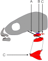
Fig 1. Schematic of the pharyngeal jaw apparatus of Cichlidae, adapted from Mabuchi et al 2007 [52].
Cranium and oral jaws are shaded grey. Pharyngeal jaws are shaded red. A) The black line represents the new muscle sling connecting the lower pharyngeal jaw to the neurocranium. B) The blue area marks the location of the novel basipharyngeal joint between the upper pharyngeal jaws and the neurocranium. C) The epibranchials 4 are decoupled from the upper pharyngeal jaws. Space between the two structures indicates decoupling, and does not represent their physiological distance D) Ventral view of the lower pharyngeal jaws. The two sides have fused together along the midline.
Fig 2
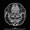
Fig 2. Transverse section of A. nigrofasciata at 6 days post fertilization.
Note the grey scale for the upper pharyngeal jaws and skull has a large range. Muscles overlap this range, preventing the use of voxel intensity to determine tissue. All structures of interest come into contact with similar intensity voxels, which causes the selection by automatic tools such as the "magic wand" to leak into adjacent areas and make them unreliable. Instead, two muscle groups (levator externus 4 and levator internus lateralis) are segmented manually here as shown by their red outlines, and this process is repeated for each structure through all images from each scan.
Fig 3
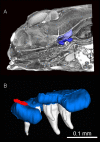
Fig 3
A) Volume rendering of A. nigrofasciata at 6 days post fertilization, lateral view. The upper pharyngeal jaws (blue), the portion of the neurocranium (light purple), the pharyngeal teeth (white) are shown as surface renderings that demonstrate their placement within the cranium. B) Finite element model of the upper pharyngeal jaws and teeth of A. nigrofasciata at 6 days post fertilization, lateral view. Jaws are light blue, teeth are white, and force vectors are the field of tightly packed red arrows. Arrows are the applied loads simulating the levator internus medialis.
Fig 4
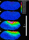
Fig 4. Comparison of muscle simulations using one node vs a set of nodes.
The left upper pharyngeal jaw is shown in the lateral view, with a portion cut away to reveal the inner stresses. Rostral is to the left, caudal to the right. A & B, First principal stress is expressed as a set of colored vectors. A) Muscle force is applied to a single node. Note the depth the higher stress values reach and how the vectors converge from both the rostral and caudal direction. B) Muscle force is applied to a set of nodes corresponding to the muscle insertion. The higher stress values are much shallower compared to 4A, and the vectors sweep further forward from the caudal end. C & D, Von Mises Stress represented by colored bricks. C) Muscle force is applied to a single node. D) Force is distributed over the area of muscle insertion. Stress at a distance from the applied force is indistinguishable in the two cases, but closer to the load in C, since the use of only one node causes exaggerated stress levels.
Fig 5
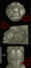
Fig 5. Reference rotational angles for the description of muscle orientation changes during ontogeny.
RCA = rostro-caudal axis. MLA = medio-lateral axis. DVA = dorso-ventral axis. Arrows indicate directions of positive angle increase.
Fig 6
Fig 6. Force on the neurocranium (NC) from the upper pharyngeal jaws in Newtons (N) in H. elegans and A. nigrofasciata.
Force in both species is comparable based on days post fertilization. Since H. elegans has a longer period with the yolk sac (17 days post fertilization) compared to A. nigrofasciata (11 days post fertilization), the former experiences a large increase in force directly after yolk sac absorption that is not seen in A. nigrofasciata.
Fig 7
Fig 7. Adduction force of the lower pharyngeal jaw from the levator externus 4 and levator posterior muscles in H. elegans and A. nigrofasciata.
While the total force always increases over time for both species, the rate of change increases between pre and post yolk sac absorption in H. elegans yet decreases during the same period in A. nigrofasciata.
Fig 8
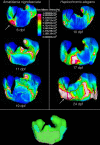
Fig 8. Locations of von Mises stress on the right upper pharyngeal jaw resulting from complete muscle contraction.
Dorsal view of the upper pharyngeal jaw of A. nigrofasciata and H. elegans. Age increases from top to bottom. Images of the upper pharyngeal jaw are not to scale, they have been adjusted in size so they can be compared. Arrows point to the area of highest stress, where the two infrapharyngobranchials attach on A. nigrofasciata at 6 days post fertilization and on H. elegans at 24 days post fertilization. The lower image in green gives an example of the placement of boundary conditions, where the UPJ met the neurocranium (pink) and the load conditions, where the muscles act on the pharyngeal jaw (blue). dpf = days post fertilization.
Fig 9
Fig 9
Dorsal view of A. nigrofasciata at (A) 6 and (B) 15 days post fertilization showing cement glands, labeled as pairs 1–3. Glands are used to attach the fry to the substrate between hatching (3 days post fertilization) and onset of free swimming (6 days post fertilization). The glands slowly decrease in size over time, but are still present at 15 days post fertilization. N = Nostril.

Similar articles

Cited by

References

    1. Heegaard JH, Beaupré GS, Carter DR. Mechanically Modulated Cartilage Growth may Regulate Joint Surface Morphogenesis. J Orthop Res. 1999;17: 509–517. doi: 10.1002/jor.1100170408 - DOI - PubMed
    1. Elder SH, Kimura JH, Soslowsky LJ, Lavagnino M, Goldstein SA. Effect of compressive loading on chondrocyte differentiation in agarose cultures of chick limb-bud cells. J Orthop Res. 2000;18: 78–86. doi: 10.1002/jor.1100180112 - DOI - PubMed
    1. van der Meulen MCH, Huiskes R. Why mechanobiology? A survey article. J Biomech. 2002;35: 401–14. - PubMed
    1. Grad S, Eglin D, Alini M, Stoddart MJ. Physical stimulation of chondrogenic cells in vitro: a review. Clin Orthop Relat Res. 2011;469: 2764–72. doi: 10.1007/s11999-011-1819-9 - DOI - PMC - PubMed
    1. Wimberger PH. Plasticity of fish body shape. The effects of diet, development, family and age in two species of Geophugus (Pisces: Cichlidae). Biol J Linn Soc. 1992;45: 197–218.

Publication types

MeSH terms

LinkOut - more resources