. 2018 Jan 10;13(1):e0190678.
doi: 10.1371/journal.pone.0190678. eCollection 2018.

Longitudinal micro-CT as an outcome measure of interstitial lung disease in TNF-transgenic mice

Affiliations

Longitudinal micro-CT as an outcome measure of interstitial lung disease in TNF-transgenic mice

Richard D Bell et al. PLoS One. .

Abstract

Introduction: Rheumatoid arthritis associated interstitial lung disease (RA-ILD) is a debilitating condition with poor survival prognosis. High resolution computed tomography (CT) is a common clinical tool to diagnose RA-ILD, and is increasingly being adopted in pre-clinical studies. However, murine models recapitulating RA-ILD are lacking, and CT outcomes for inflammatory lung disease have yet to be formally validated. To address this, we validate μCT outcomes for ILD in the tumor necrosis factor transgenic (TNF-Tg) mouse model of RA.

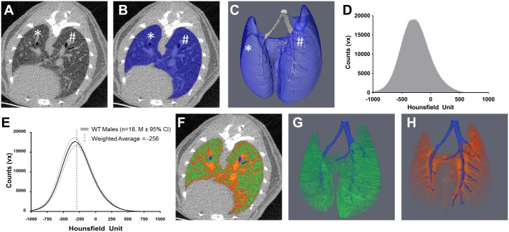
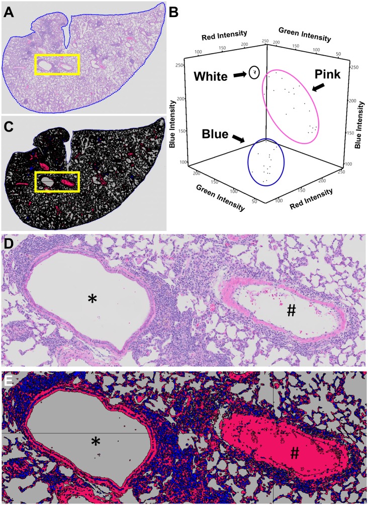
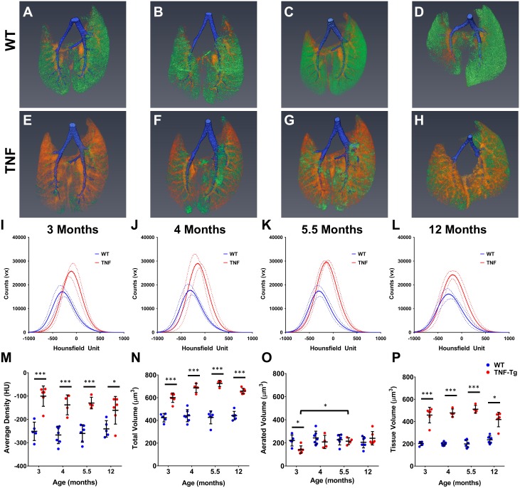
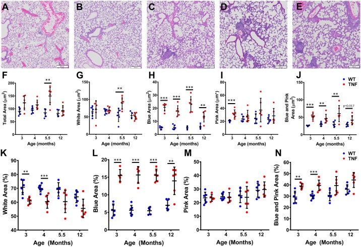
Methods: Cross sectional μCT was performed on cohorts of male TNF-Tg mice and their WT littermates at 3, 4, 5.5 and 12 months of age (n = 4-6). Lung μCT outcomes measures were determined by segmentation of the μCT datasets to generate Aerated and Tissue volumes. After each scan, lungs were obtained for histopathology and 3 sections stained with hematoxylin and eosin. Automated histomorphometry was performed to quantify the tissue area (nuclei, cytoplasm, and extracellular matrix) and aerated area (white space) within the tissue sections. Spearman's correlation coefficients were used to evaluate the extent of association between μCT imaging and histopathology endpoints.

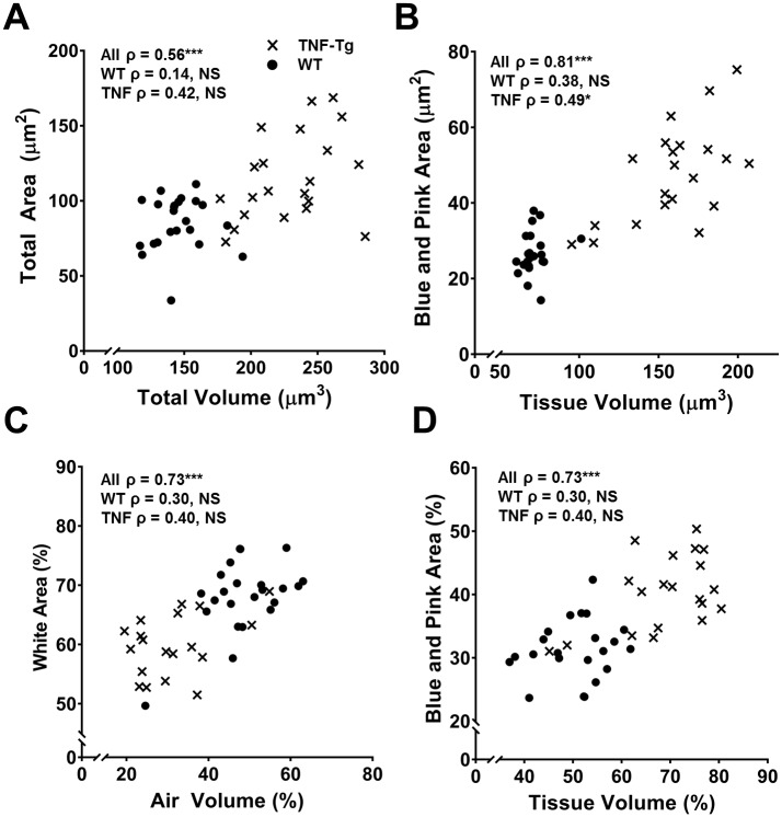
Results: TNF-Tg mice had significantly greater tissue volume, total lung volume and mean intensity at all timepoints compared to age matched WT littermates. Histomorphometry also demonstrated a significant increase in tissue area at 3, 4, and 5.5 months of age in TNF-Tg mice. Lung tissue volume was correlated with lung tissue area (ρ = 0.81, p<0.0001), and normalize lung aerated volume was correlated with normalized lung air area (ρ = 0.73, p<0.0001).

Conclusions: We have validated in vivo μCT as a quantitative biomarker of ILD in mice. Further, development of longitudinal measures is critical for dissecting pathologic progression of ILD, and μCT is a useful non-invasive method to study lung inflammation in the TNF-Tg mouse model.


Conflict of interest statement

Competing Interests: The authors have declared that no competing interests exist.

Figures

Fig 1
Fig 1. In vivo micro-CT quantification of aerated and tissue lung volume.
A) A 2D representative μCT transverse, inferior cross section of a WT lung at cervical vertebra 8 shows the margin of the lung inside of the abdominal cavity. B) This margin is then used with semi-automated tools in Amira to segment the lung (Blue overlay) and a 3D reconstruction can be generated (C). The lung is further segmented into the conducting airway (Grey, C) and into the left (*) and right (#) lobes for more specific analysis (A-C). The raw intensity values from either the whole lung segment or a smaller portion are extracted to generate intensity histograms (D). To delineate aerated lung volume from tissue lung volume a thresholding operation must be developed. The most abundant volume in a healthy lung is the interface between air and the alveolar epithelium. As measured by μCT, the most abundant density in 3–12 month old WT mice is -256 HU (E). Using this threshold, aerated and tissue lung volume can be quantified as shown in a representative 2D overlay (F, Green = Aerated Volume, Orange-Red = Tissue Volume, Blue = Conducting Airway) and 3D reconstructions (G, Aerated Volume; H, Tissue Volume, Blue = Conducting Airway). Note the distinct bronchiole and arteriole structures in G and H.
Fig 2
Fig 2. Histomorphometry method for identifying cell nuclei, cytoplasm, extracellular matrix, red blood cells and alveolar space with Visiopharm.
To quantify histomorphometric areas, lungs were stained with Hematoxylin and Eosin, whole slides were scanned, imported into Visiopharm and a ROI was drawn around the margin of the lung (A, ROI = blue dotted line). A custom application was developed to classify the assorted colors within the ROI of the stained slide into 3 categories (Blue, Pink, and White) by training Visiopharm on the Red/Green/Blue pixel intensity ranges. This occurs through manual selection of a training data set of RGB values for each category and application of a Bayesian discrimination algorithm to determine a set of RGB ranges for each category. An example training data set with >15 pixels for each category is presented in B with the intensity of the Red, Blue and Green pixels plotted against each other with each category circled together in 3D space. Note how each category has a distinct range of intensities allowing discrimination of cell nuclei (Blue category) from cytoplasm, ECM and RBCs (Pink category) from alveolar space (White category). Once defined, this classification was applied to every slide in the data set. Representative H and E (A and D) and classified slides (C and E) are shown to demonstrate the specificity of our classification method (bronchiole = *; artery = #). For arteries and arterioles that were not filled with RBCs, manual classification was performed to change the space from White to Pink after inspection of the epithelial layer (#, E).
Fig 3
Fig 3. TNF-Tg male mice have increased tissue volume compared to WT littermates measured via μCT.
Representative 3D reconstructions of 3 (A, E), 4 (B, F), 5.5 (C, G), and 12 (D, H) month old WT and TNF-Tg male mice show a clear increase in tissue volume (Orange-Red) in the TNF-Tg animals at all timepoints while maintaining a similar amount of aerated volume (Green) at all timepoints. Conducting airways that were segmented out of analysis shown in Blue. Histograms of the extracted data from the whole lung segmentation for each timepoint is presented in I-L (M ± 95%CI, n = 4–6). TNF-Tg male mice have a statistically significant increase in mean lung intensity (M), total lung volume (N) and tissue lung volume (P) at all timepoints compared WT littermates (*p<0.05, ***p<0.001, M±SD, n = 4–6).
Fig 4
Fig 4. TNF-Tg male mice have greater tissue area compared to WT litter mates measured via histomorphometry.
Blue, Pink and White areas were measured on 3 sections >200 μm apart for 3, 4, 5.5, and 12-month-old male TNF-Tg and WT littermates. Representative H and E images from a 12-month WT (A), and 3 (B), 4 (C), 5 (D) and 12 (E) month TNF-Tg mice show a clear increase in cells in the TNF-Tg mice at all timepoints. Quantifying each histomorphometric category demonstrates a significant increase in Total area at 3 and 5.5 months (F), Blue area at all timepoints (G) and Blue + Pink at 3, 4, and 5.5 months (H) for TNF-Tg mice compared to their WT littermates. Due to the known volumetric changes that occur in the histologic processing solutions (i.e. formalin, ethanols) and the nature of lung tissue being sensitive to changes due to its composition, we normalized all the measurements to the total area (I, J, K and L). As expected, percent Blue area (J) at all timepoints and percent Blue + Pink (L) at 3, 4 and 5.5 months were significantly increased in TNF-Tg mice compared to WT littermates. Interestingly, when normalized to total area, there was a significant decrease in the White area at 3, 4, and 5.5 months in TNF-Tg mice compared to WT littermates (K; *p<0.0125, **p<0.01, ***p<0.001, M±SD, n = 4–6).
Fig 5
Fig 5. Tissue volume and tissue area (Blue + Pink) are highly correlated.
Spearman’s correlations were performed comparing μCT outcome measures and histomorphometry outcome measures. Both total volume and total area (A) as well as tissue volume and Blue + Pink (C) area were highly correlated with each other. Aerated volume and White area were not correlated (Not Shown); however, when normalized to total volume and area (C), there was a significant correlation. Normalized tissue volume and Blue + Pink area remained correlated (D). Spearman Rank coefficients are present on each graph for all data points (All ρ) and for each genotype independently (WT ρ, TNF-Tg ρ, X = TNF-Tg, ● = WT, n = 43, NS = not significant, *p<0.05, ***p<0.0001).

References

    1. Bongartz T, Nannini C, Medina-Velasquez YF, Achenbach SJ, Crowson CS, Ryu JH, et al. Incidence and mortality of interstitial lung disease in rheumatoid arthritis: a population-based study. Arthritis Rheum. 2010;62(6):1583–91. doi: 10.1002/art.27405 - DOI - PMC - PubMed
    1. Doyle JJ, Eliasson AH, Argyros GJ, Dennis GJ, Finger DR, Hurwitz KM, et al. Prevalence of pulmonary disorders in patients with newly diagnosed rheumatoid arthritis. Clin Rheumatol. 2000;19(3):217–21. - PubMed
    1. Kim EJ, Elicker BM, Maldonado F, Webb WR, Ryu JH, Van Uden JH, et al. Usual interstitial pneumonia in rheumatoid arthritis-associated interstitial lung disease. Eur Respir J. 2010;35(6):1322–8. doi: 10.1183/09031936.00092309 - DOI - PubMed
    1. Shaw M, Collins BF, Ho LA, Raghu G. Rheumatoid arthritis-associated lung disease. Eur Respir Rev. 2015;24(135):1–16. doi: 10.1183/09059180.00008014 - DOI - PMC - PubMed
    1. Raghu G, Collard HR, Egan JJ, Martinez FJ, Behr J, Brown KK, et al. An official ATS/ERS/JRS/ALAT statement: idiopathic pulmonary fibrosis: evidence-based guidelines for diagnosis and management. Am J Respir Crit Care Med. 2011;183(6):788–824. doi: 10.1164/rccm.2009-040GL - DOI - PMC - PubMed

Publication types