. 2018 Jan 9;27(1):252-262.e3.
doi: 10.1016/j.cmet.2017.12.004.

An Adipose Tissue Atlas: An Image-Guided Identification of Human-like BAT and Beige Depots in Rodents

Affiliations

An Adipose Tissue Atlas: An Image-Guided Identification of Human-like BAT and Beige Depots in Rodents

Fang Zhang et al. Cell Metab. .

Abstract

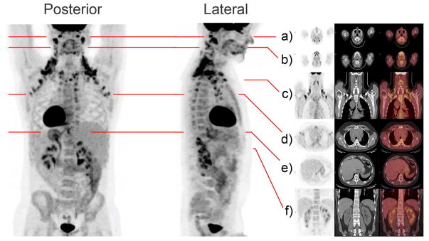
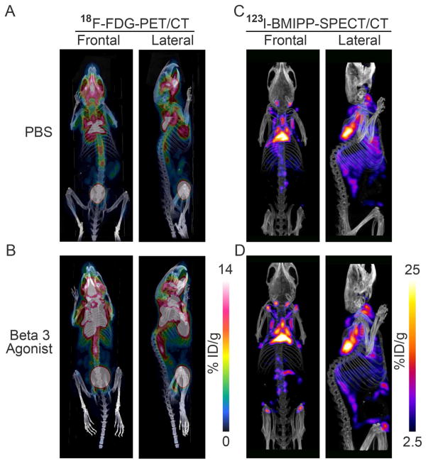
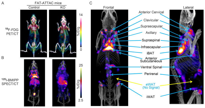
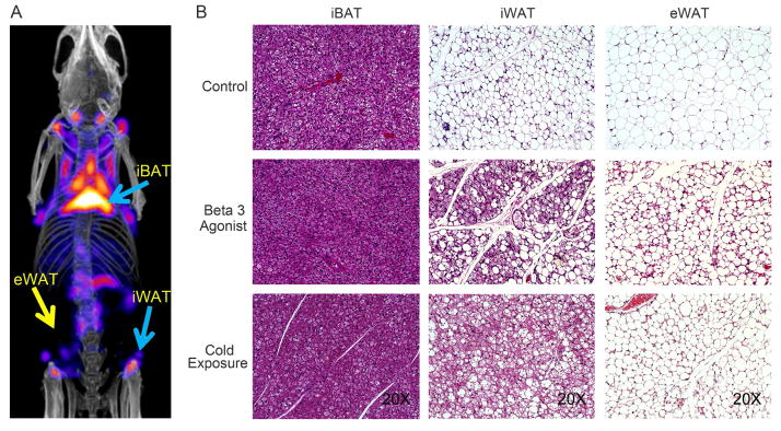
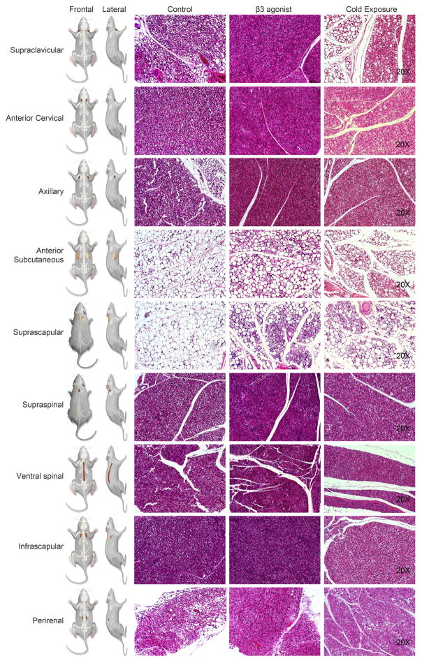
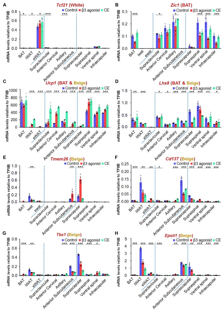
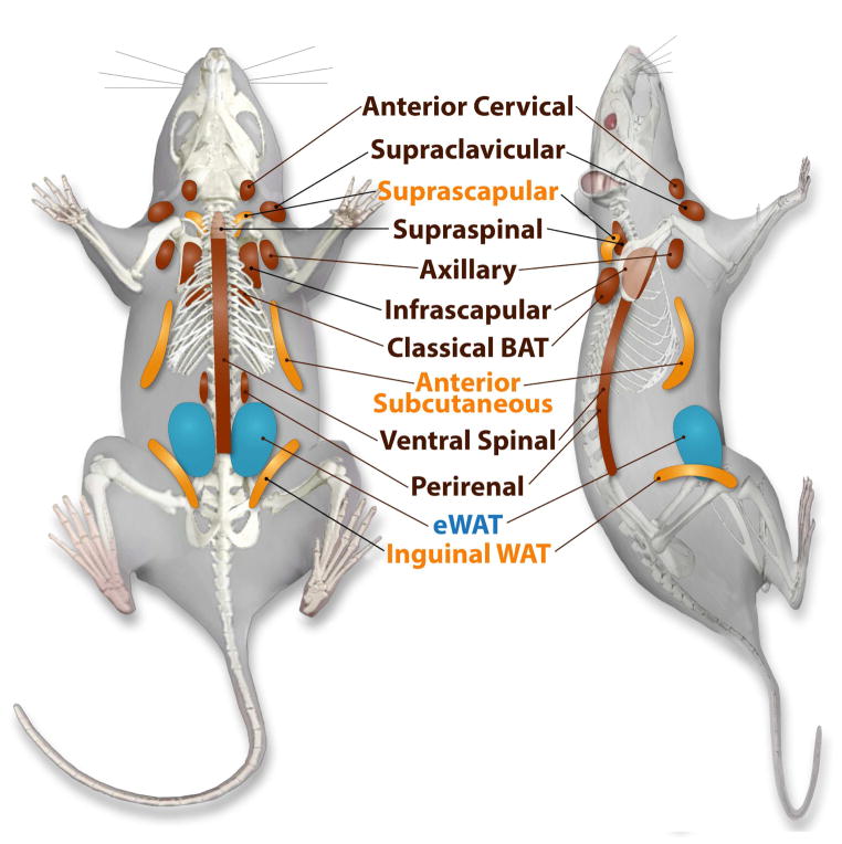
[18F]Fluorodeoxyglucose-PET/CT (18F-FDG-PET/CT) imaging has been invaluable for visualizing metabolically active adipose tissues in humans with potential anti-diabetic and anti-obesity effects. To explore whether mice display human-like fat depots in anatomically comparable regions, we mapped fat depots using glucose or fatty acid imaging tracers, such as 18F-FDG through PET/CT or [123/125I]-β-methyl-p-iodophenyl-pentadecanoic acid with SPECT/CT imaging, to analogous depots in mice. Using this type of image analysis with both probes, we define a large number of additional areas of high metabolic activity corresponding to novel fat pads. Histological and gene expression analyses validate these regions as bona fide fat pads. Our findings indicate that fat depots of rodents show a high degree of topological similarity to those of humans. Studies involving both glucose and lipid tracers indicate differential preferences for these substrates in different depots and also suggest that fatty acid-based visualized approaches may reveal additional brown adipose tissue and beige depots in humans.

Keywords: BAT; PET/CT; SPECT/CT; beige; bona fide thermogenic fat tissues; glucose or fatty acid imaging tracers; human-like; imaging; marker genes; morphology.


Conflict of interest statement

Conflicts of interest: none.

Figures

Figure 1
Figure 1. Regions of High Level Glucose Uptake in Humans as Assessed by 18F-FDG-PET/CT
Metabolic activity in thermogenic tissues is demonstrated in this subject imaged with integrated [18F]-Fluorodeoxyglucose-Positron Emission Tomography–Computed Tomography (18F-FDG-PET/CT) at room temperature. Frontal and lateral maximal PET intensity projection images in an adult male subject show distribution of glucose uptake by its image tracer 18F-FDG. In this subject, prominent FDG retention is shown in the cervical, supraclavicular, axillary, intercostal, mediastinal, ventral spinal, and peri-renal areas. Paravertebral tissue is also activated. Fat pads are also illustrated in multi-level transaxial or coronal PET/CT images (a–f) are shown in the Supplemental Movie (Movie S1).
Figure 2
Figure 2. Anatomical Location of Metabolically Active Fat Pads as Assessed by 3D-images of PET/CT with 18F-FDG for Glucose Uptake and SPECT/CT with 123I-BMIPP for Fatty Acid Uptake
A-B. The left panels show the 18F-FDG-PET/CT images of glucose uptake and distribution in the thermogenic tissues of an adult mouse kept at room temperature treated with 7-days PBS (upper-left panels, A) or β3 agonist (down-left panels, B). C–D. The right panels show the 123I-BMIPP-SPECT/CT scanning for the patterns of fatty acid uptake in the metabolic active tissues of an adult mouse kept at room temperature after 7-days PBS treatment (upper-right panels, C) or β3 adrenergic stimulation. (lower-right panels, D). In each case, representative images for n=3 repeats are shown.
Figure 3
Figure 3. Signal intensity in a Lipodystrophic Rodent Model
A. A model of inducible lipodystrophy (the “FAT-ATTAC mouse”) selectively loses adipocyte-derived signals upon 18F-FDG injection compared to control. B. SPECT/CT imaging of the FAT-ATTAC lipodystrophic mouse confirms the identity of signals as adipocyte-derived 125I-BMIPP uptake. C. The main metabolically active adipose tissues indicated with blue arrows in an adult mouse model stimulated by β3-adrenergic agonist for 7 days. Blue color label with yellow arrows indicate fat pads that display background signal levels of 18F-FDG-PET/CT and 123I-BMIPP-SPECT/CT. In each case, representative images for n=3 repeats are shown.
Figure 4
Figure 4. Histological Analysis of interscapular BAT, inguinal and epididymal fat tissues
A. Locations of interscapular BAT (iBAT), inguinal WAT (iWAT) and epididymal WAT (eWAT) depots indicated with blue arrows or yellow arrows display differential levels of fatty acid uptake after 7-days β3 adrenergic stimulation. B. Histological analysis of tissues highlights the expected phenotype of these “classic” adipose tissues from control, β3 agonist - treated and cold-stimulated mice. The presented images are representative for n=4 repeats of the histological analysis.
Figure 5
Figure 5. Histological Analysis of Additional Fat Pads at Baseline, β3 Agonist – Treated and Cold-Stimulated Mice
The images explore a series of additional regions of high metabolic activity around supraclavicular, anterior cervical, axillary, anterior subcutaneous, suprascapular, supraspinal, ventral spinal, infrascapular regions and in the peri-renal area as indicated in the cartoon. Clearly apparent are the unilocular resp. multilocular characteristics of each of the fat pads of control, β3 agonist-treated and cold-stimulated mice. The presented images are representative for n=4 repeats of the histological analysis
Figure 6
Figure 6. Expression Levels of Marker Genes Classify the Newly Isolated Fat Pads into the Three Categories of Fat Pads including WAT, BAT and Beige
A series of marker genes including Tcf21 for WAT, Zic1 for BAT depots, Ucp1 and Lhx8 for BAT and beige depots and Tmem26, Cd137, Tbx1 and Epsti1 for beige depots were used to examine the mRNA expression patterns of the newly identified fat pads, including supraclavicular, anterior cervical, axillary, anterior subcutaneous, suprascapular, supraspinal, ventral spinal, infrascapular regions and in the perirenal area in mice under control, β3 agonist-treated and cold-stimulated conditions (n=6). Results are shown as mean ± SD, *p <0.05, **p < 0.01 and ***p < 0.001.
Figure 7
Figure 7. Location of Metabolically Identified and Histologically Confirmed Fat Pads
White adipose tissues indicated in blue, fat pads with the ability to beige are in yellow, and more classical brown adipose tissue-like fat pads are indicated in brown.

Similar articles

Cited by

References

    1. Admiraal WM, Holleman F, Bahler L, Soeters MR, Hoekstra JB, Verberne HJ. Combining 123I-metaiodobenzylguanidine SPECT/CT and 18F-FDG PET/CT for the assessment of brown adipose tissue activity in humans during cold exposure. J Nucl Med. 2013;54:208–212. - PubMed
    1. Betz MJ, Enerback S. Human Brown Adipose Tissue: What We Have Learned So Far. Diabetes. 2015;64:2352–2360. - PubMed
    1. Cannon B, Nedergaard J. Brown adipose tissue: function and physiological significance. Physiol Rev. 2004;84:277–359. - PubMed
    1. Ci X, Frisch F, Lavoie F, Germain P, Lecomte R, van Lier JE, Benard F, Carpentier AC. The effect of insulin on the intracellular distribution of 14(R,S)-[18F]Fluoro-6-thia-heptadecanoic acid in rats. Mol Imaging Biol. 2006;8:237–244. - PubMed
    1. Cinti S. Transdifferentiation properties of adipocytes in the adipose organ. Am J Physiol Endocrinol Metab. 2009;297:E977–986. - PubMed

Publication types

MeSH terms

LinkOut - more resources