Case Reports
. 2019 May:128:140-149.
doi: 10.1016/j.neuropsychologia.2018.01.007. Epub 2018 Jan 7.

Subcortical pathways to extrastriate visual cortex underlie residual vision following bilateral damage to V1

Affiliations
Case Reports

Subcortical pathways to extrastriate visual cortex underlie residual vision following bilateral damage to V1

Sara Ajina et al. Neuropsychologia. 2019 May.

Abstract

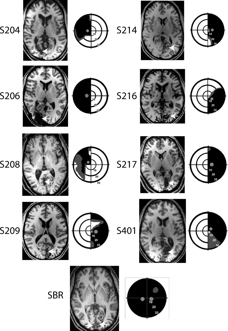
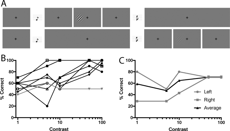
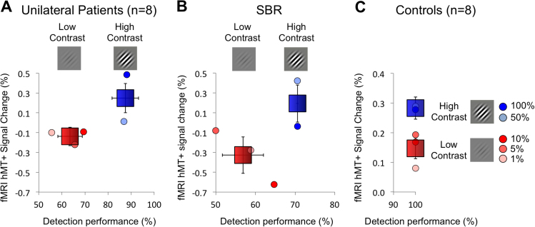
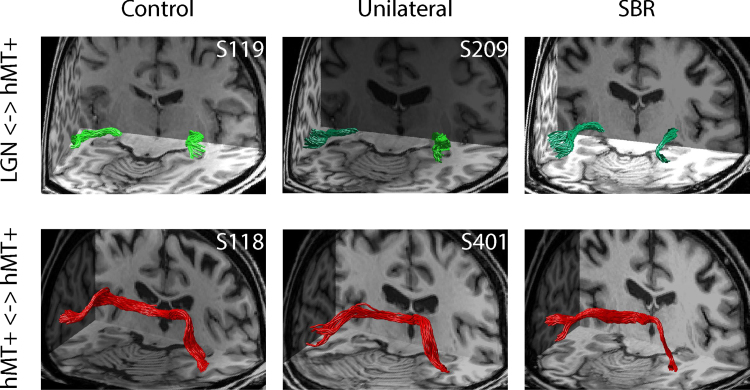
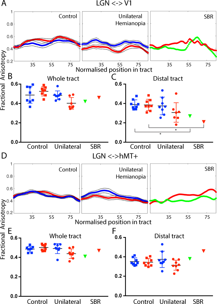
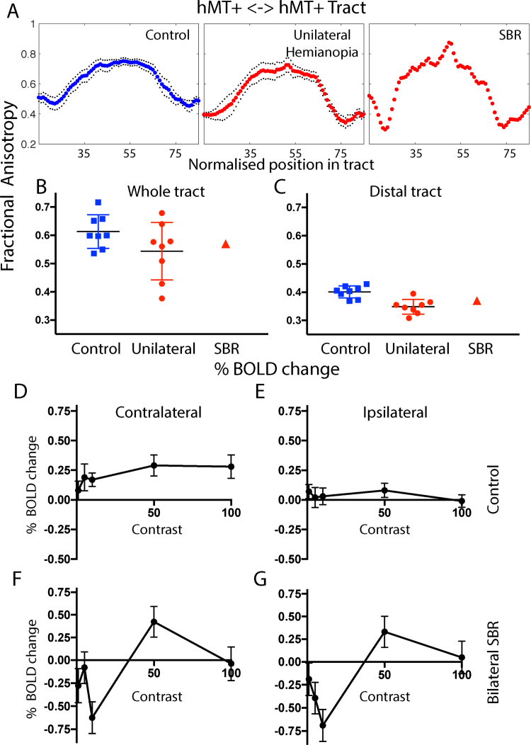
Residual vision, or blindsight, following damage to the primary visual cortex (V1) has been investigated for almost half a century. While there have been many studies of patients with unilateral damage to V1, far fewer have examined bilateral damage, mainly due to the rarity of such patients. Here we re-examine the residual visual function and underlying pathways of previously studied patient SBR who, as a young adult, suffered bilateral damage restricted to V1 which rendered him cortically blind. While earlier work compared his visual cortex to healthy, sighted participants, here we consider how his visual responses and connections compare to patients with unilateral damage to V1 in addition to sighted participants. Detection of drifting Gabor patches of different contrasts (1%, 5%, 10%, 50% and 100%) was tested in SBR and a group of eight patients with unilateral damage to V1. Performance was compared to the neural activation in motion area hMT+ measured using functional magnetic resonance imaging. Diffusion tractography was also used to determine the white matter microstructure of the visual pathways in all participants. Like the patients with unilateral damage, patient SBR showed increased % BOLD signal change to the high contrast stimuli that he could detect compared to the lower contrast stimuli that were not detectable. Diffusion tractography suggests this information is conveyed by a direct pathway between the lateral geniculate nucleus (LGN) and hMT+ since this pathway had microstructure that was comparable to the healthy control group. In contrast, the pathway between LGN and V1 had reduced integrity compared to controls. A further finding of note was that, unlike control participants, SBR showed similar patterns of contralateral and ipsilateral activity in hMT+, in addition to healthy white matter microstructure in the tract connecting hMT+ between the two hemispheres. This raises the possibility of increased connectivity between the two hemispheres in the absence of V1 input. In conclusion, the pattern of visual function and anatomy in bilateral cortical damage is comparable to that seen in a group of patients with unilateral damage. Thus, while the intact hemisphere may play a role in residual vision in patients with unilateral damage, its influence is not evident with the methodology employed here.

Keywords: Cortical blindness; Hemianopia; V1; Visual perception.


Figures

Fig. 1
Fig. 1
The lesion and visual field loss is shown for each patient with unilateral hemianopia and SBR. The arrows indicate the location of the lesion for each patient. The visual fields are presented schematically based upon Humphrey perimetry, with black indicating most severe visual loss and grey indicating partial loss. The visual fields of S216 are based on Goldmann perimetry. The small Gabor patches indicate the location in which the visual stimuli were presented to the patient.
Fig. 2
Fig. 2
A shows the psychophysical task performed by the patients with unilateral homonymous hemianopia (n = 8) and patient SBR who has bilateral cortical blindness. The patients were required to determine which of the two intervals contained the Gabor patch. The upper row shows a high contrast Gabor patch, while the lower row shows a low contrast Gabor patch. In each case the Gabor patch is only presented for 500 ms rather than the entire trial. B shows the performance of each of the unilateral patients at the different contrast levels. Only one patient (grey line) was unable to perform this task above chance when the stimulus was at 100% contrast. C shows the performance of SBR in the two hemifields, and indicates his ability to perform above chance at the higher two contrasts.
Fig. 3
Fig. 3
In the blind hemifield of the 8 patients with unilateral damage to V1 and in patient SBR with bilateral V1 damage, the %BOLD change in hMT+ to high contrast stimuli (50% and 100%) is significantly higher than that to low contrasts (1%, 5%, 10%). Healthy control participants (n=8) also show a higher response to the high contrast stimuli, but performance would be at ceiling for both high and low contrast detection (note the different scale on both axes). Signal change and performance scores are also provided for each contrast level separately.
Fig. 4
Fig. 4
Tracts between LGN and hMT+ were identified in control participants, patients with unilateral hemianopia and SBR (upper row). Tracts in healthy hemisphere are shown in light green, whereas those in the damaged hemisphere are dark green. Tracts between hMT+ in the two hemispheres were also determined (lower row), shown in red. (For interpretation of the references to color in this figure legend, the reader is referred to the web version of this article.)
Fig. 5
Fig. 5
The upper section (panels A–C) shows the fractional anisotropy (FA) in the tract between LGN and V1. A shows the FA along the length of the tract from LGN to V1 for control participants (red: left hemisphere, blue: right hemisphere), patients with unilateral V1 damage (red: lesion hemisphere, blue: intact hemisphere) and SBR (red: left hemisphere, green: right hemisphere). B is the average FA across the whole tract for each group and hemisphere. C shows the average FA in the distal portion of the tract (nodes 85–100). The asterisk indicates that SBR's FA values are ≥ 2 sd. below the mean control value. (For interpretation of the references to color in this figure legend, the reader is referred to the web version of this article.)
Fig. 6
Fig. 6
Communication between the two hemispheres may be important in residual vision. The microstructure of the connection between hMT+ in the two hemispheres is intact in SBR, with relatively high FA (A-C). In control participants there is a clear pattern of response to contrast in the contralateral hMT+ that saturates at low values. The ipsilateral response is considerably lower and shows a less clear pattern. Interestingly, in SBR the contralateral and ipsilateral responses to visual stimulation are very similar. Error bars show standard deviations.

References

    1. Ajina S., Kennard C., Rees G., Bridge H. Motion area V5/MT+ response to global motion in the absence of V1 resembles early visual cortex. Brain. 2015;138:164–178. - PMC - PubMed
    1. Ajina S., Pestilli F., Rokem A., Kennard C., Bridge H. Human blindsight is mediated by an intact geniculo-extrastriate pathway. Elife. 2015:4. - PMC - PubMed
    1. Ajina S., Rees G., Kennard C., Bridge H. Abnormal contrast responses in the extrastriate cortex of blindsight patients. J. Neurosci. 2015;35:8201–8213. - PMC - PubMed
    1. Allen B., Spiegel D.P., Thompson B., Pestilli F., Rokers B. Altered white matter in early visual pathways of humans with amblyopia. Vision. Res. 2015;114:48–55. - PubMed
    1. Andersson J. L. R., Jenkinson M., Smith S. (2007). Non-linear registration aka spatial normalisation. Technical Report FMRIB Technical Report TR07JA2, Oxford: FMRIB Centre.

Publication types

MeSH terms