. 2018 Jan 9;22(2):332-339.
doi: 10.1016/j.celrep.2017.12.060.

Esrrb Complementation Rescues Development of Nanog-Null Germ Cells

Affiliations

Esrrb Complementation Rescues Development of Nanog-Null Germ Cells

Man Zhang et al. Cell Rep. .

Abstract

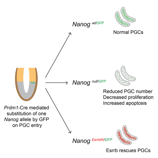
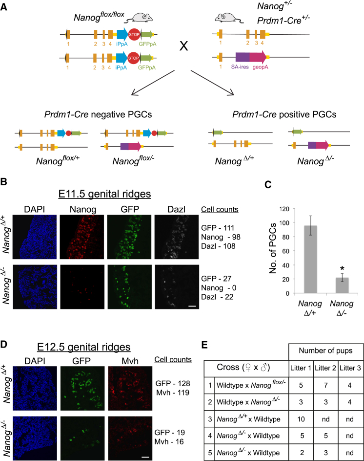
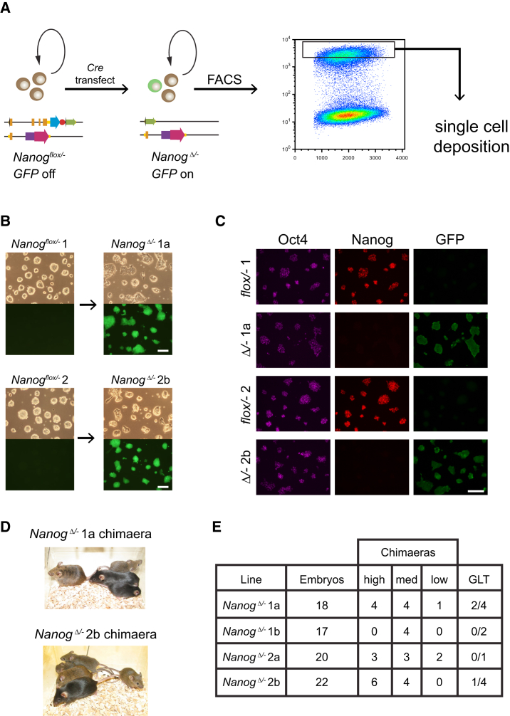
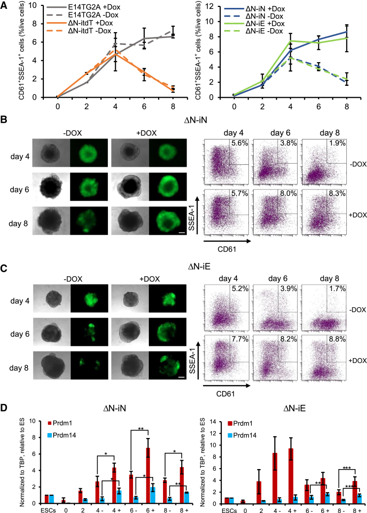
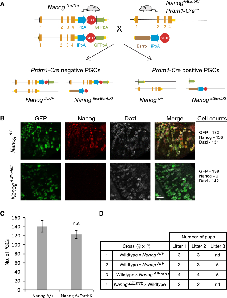
The transcription factors (TFs) Nanog and Esrrb play important roles in embryonic stem cells (ESCs) and during primordial germ-cell (PGC) development. Esrrb is a positively regulated direct target of NANOG in ESCs that can substitute qualitatively for Nanog function in ESCs. Whether this functional substitution extends to the germline is unknown. Here, we show that germline deletion of Nanog reduces PGC numbers 5-fold at midgestation. Despite this quantitative depletion, Nanog-null PGCs can complete germline development in contrast to previous findings. PGC-like cell (PGCLC) differentiation of Nanog-null ESCs is also impaired, with Nanog-null PGCLCs showing decreased proliferation and increased apoptosis. However, induced expression of Esrrb restores PGCLC numbers as efficiently as Nanog. These effects are recapitulated in vivo: knockin of Esrrb to Nanog restores PGC numbers to wild-type levels and results in fertile adult mice. These findings demonstrate that Esrrb can replace Nanog function in germ cells.

Keywords: PGCLCs; competence; naive pluripotency; primordial germ cells; transcription factors.


Figures

None
Graphical abstract
Figure 1
Figure 1
Conditional Deletion of Nanog Reduces PGC Numbers (A) Strategy for Nanog conditional knockout. Nanogflox/flox females are crossed with Nanog+/−; Prdm1-Cre male mice. As Prdm1-Cre is heterozygous, one in four embryos will have germline deletion of Nanog (NanogΔ/−). (B) E11.5 genital ridge sections from NanogΔ/− and control embryos immunostained for Nanog, Dazl, and GFP and counterstained with 4',6-diamidino-2-phenylindole (DAPI) (scale bar, 50 μm). (C) Cell counts of PGCs in NanogΔ/− and control genital ridges at E11.5. PGCs identified by co-staining for Oct4 and either Dazl or Mvh. The mean (± SD) of two biological and technical replicates for each sample are shown. p < 0.05 (unpaired Student’s t test). (D) E12.5 genital ridges from NanogΔ/− and control embryos immunostained for GFP and Mvh and counterstained with DAPI (scale bar, 50 μm). (E) Table of breeding data for adult NanogΔ/− mice. Both male (row 2) and female (row 4 and 5) NanogΔ/− mice are fertile. See also Figure S1.
Figure 2
Figure 2
Contribution of Nanog-Null ESCs to Adult Chimeras, Including the Germline (A) Strategy for generation NanogΔ/− (Nanog-null) clonal ESC lines. (B) Phase contrast and fluorescence images of parental and Nanog-null ESC lines (scale bar, 100 μm). (C) Oct4, Nanog, and GFP immunostaining of parental and Nanog-null ESC lines (scale bar, 100 μm). (D) Chimeras generated from Nanog-null ESCs, C57BL/6 mates, and agouti and black pups. High-contribution chimeras generated by injection of agouti Nanog-null ESCs into C57BL/6 blastocysts. (E) Summary of blastocyst injections and germline contribution of four clonal Nanog-null ESC lines. See also Figures S2 and S3.
Figure 3
Figure 3
Esrrb Can Replace the Nanog Requirement for Efficient PGCLC Differentiation (A) The proportion of SSEA1+/CD61+ cells during PGC differentiation of E14TG2A and ΔN-itdT (left) or ΔN-iNanog (ΔN-iN) and ΔN-iEsrrb (ΔN-iE) (right) ESCs are shown at the indicated days of PGCLC differentiation in the absence (−) or presence (+) of Dox addition from day 2 onward (please refer to Figure S5A for differentiation protocol details). Values are means ± SDs; n = 3 biological replicates. (B and C) PGCLC differentiation of ΔN-iN (B) and ΔN-iE (C) ESCs in the presence (+) or absence (−) of Dox. The morphology and Nanog:GFP expression of aggregates are shown (left; scale bar, 200 μm) with SSEA1/CD61 analysis by fluorescence-activated cell sorting (FACS) (right). (D) Quantitative mRNA expression analysis during PGC differentiation of ΔN-iN (left) and ΔN-iE (right) in the presence (+) or absence (−) of Dox at the indicated number of days of PGCLC differentiation. Values are means ± SDs; n = 3 biological replicates. p < 0.05; ∗∗p < 0.01; and ∗∗∗p < 0.001 (unpaired Student’s t test). See also Figures S4–S6.
Figure 4
Figure 4
Esrrb Expression Can Rescue Development of Nanog−/− PGCs (A) Schematic of Nanog conditional knockout, Esrrb knockin strategy. Nanogflox/flox female mice are crossed with Prdm1-Cre: Nanog+/EsrrbKI male mice. As Prdm1-Cre is heterozygous, one in four offspring will be Nanog conditional knockout, Esrrb knockin (NanogΔ/EsrrbKI). (B) E12.5 genital ridges from NanogΔ/EsrrbKI and control embryos. GFP expression is from the conditionally deleted (Δ) allele and is specific to germ cells of the genital ridge (scale bar, 50 μm). (C) Cell numbers were counted from NanogΔ/EsrrbKI and control genital ridges. PGCs are identified by Dazl expression. The mean (± SD) of three biological replicates for control and NanogΔ/EsrrbKI are shown. n.s., not significant. (D) Table of breeding data for adult NanogΔ/EsrrbKI and control mice. Both male (row 3) and female (row 4) NanogΔ/EsrrbKI mice are fertile. See also Figures S7 and S8.

References

    1. Beddington R.S. An autoradiographic analysis of tissue potency in different regions of the embryonic ectoderm during gastrulation in the mouse. J. Embryol. Exp. Morphol. 1982;69:265–285. - PubMed
    1. Boroviak T., Loos R., Bertone P., Smith A., Nichols J. The ability of inner-cell-mass cells to self-renew as embryonic stem cells is acquired following epiblast specification. Nat. Cell Biol. 2014;16:516–528. - PMC - PubMed
    1. Brook F.A., Gardner R.L. The origin and efficient derivation of embryonic stem cells in the mouse. Proc. Natl. Acad. Sci. USA. 1997;94:5709–5712. - PMC - PubMed
    1. Campolo F., Gori M., Favaro R., Nicolis S., Pellegrini M., Botti F., Rossi P., Jannini E.A., Dolci S. Essential role of Sox2 for the establishment and maintenance of the germ cell line. Stem Cells. 2013;31:1408–1421. - PubMed
    1. Carter A.C., Davis-Dusenbery B.N., Koszka K., Ichida J.K., Eggan K. Nanog-independent reprogramming to iPSCs with canonical factors. Stem Cell Reports. 2014;2:119–126. - PMC - PubMed

Publication types