Review
. 2017 Dec;55(6):587-599.
doi: 10.3347/kjp.2017.55.6.587. Epub 2017 Dec 31.

Therapeutic Effects of Resiniferatoxin Related with Immunological Responses for Intestinal Inflammation in Trichinellosis

Affiliations
Review

Therapeutic Effects of Resiniferatoxin Related with Immunological Responses for Intestinal Inflammation in Trichinellosis

José Luis Muñoz-Carrillo et al. Korean J Parasitol. 2017 Dec.

Abstract

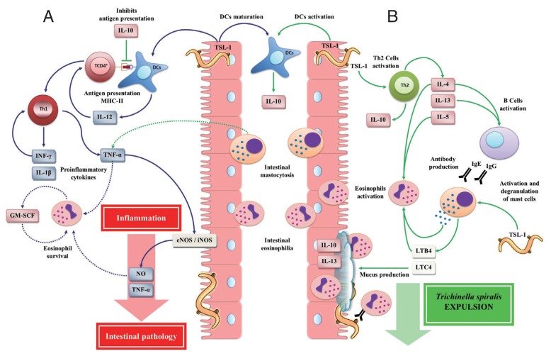
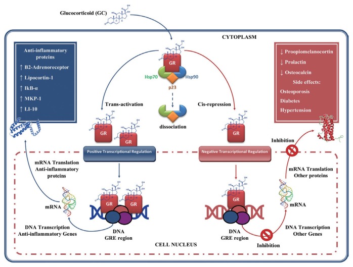
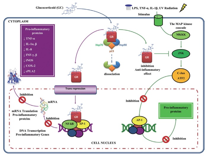
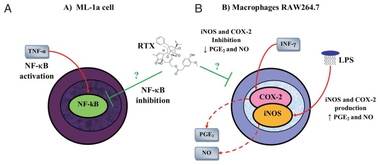
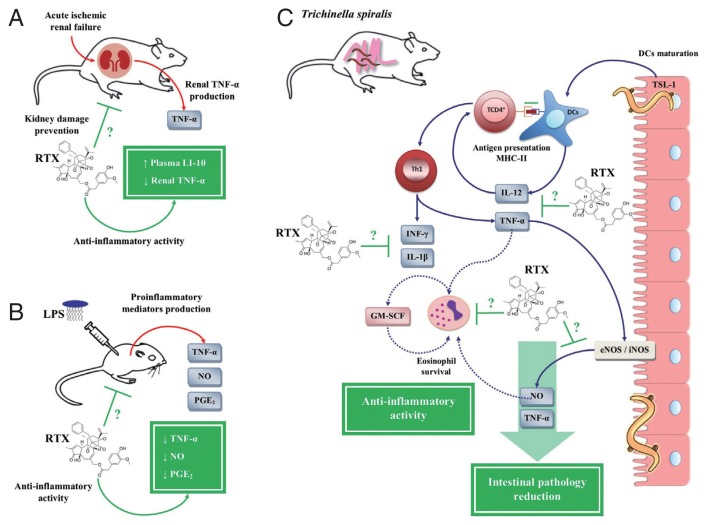
The immune response against Trichinella spiralis at the intestinal level depends on the CD4+ T cells, which can both suppress or promote the inflammatory response through the synthesis of diverse cytokines. During the intestinal phase, the immune response is mixed (Th1/Th2) with the initial predominance of the Th1 response and the subsequent domination of Th2 response, which favor the development of intestinal pathology. In this context, the glucocorticoids (GC) are the pharmacotherapy for the intestinal inflammatory response in trichinellosis. However, its therapeutic use is limited, since studies have shown that treatment with GC suppresses the host immune system, favoring T. spiralis infection. In the search for novel pharmacological strategies that inhibit the Th1 immune response (proinflammatory) and assist the host against T. spiralis infection, recent studies showed that resiniferatoxin (RTX) had anti-inflammatory activity, which decreased the serum levels of IL-12, INF-γ, IL-1β, TNF-α, NO, and PGE2, as well the number of eosinophils in the blood, associated with decreased intestinal pathology and muscle parasite burden. These researches demonstrate that RTX is capable to inhibit the production of Th1 cytokines, contributing to the defense against T. spiralis infection, which places it as a new potential drug modulator of the immune response.

Keywords: Th1 cytokine; Trichinella spiralis; inflammatory response; resiniferatoxin; trichinellosis.


Conflict of interest statement

CONFLICT OF INTEREST

We have no conflict of interest related to this work.

Figures

Fig. 1
Fig. 1
Life cycle of Trichinella spiralis. 1) Infected meat ingestion with T. spiralis. Intestinal phase. 2) Release of infective larvae of T. spiralis (T. spiralis-L1) in the stomach. 3) Migration of T. spiralis-L1 to the small intestine and maturation to female and male adult worms (AD). 4) Reproduction and release of newborn larvae (NBL) of T. spiralis. Muscle phase. 5) Migration of NBL T. spiralis and invasion of skeletal muscle cells to develop to a stage of T. spiralis-L1 forming the nurse cell (NC). This figure was made by the authors based on the references cited in the text.
Fig. 2
Fig. 2
Immune responses during the intestinal phase of T. spiralis infection. (A) T helper type 1 (Th1) immune response: T. spiralis larvae group antigens (TSL-1) induce maturation of dendritic cells (DCs) by polarizing a Th1 immune response, which is mainly characterized by the release of interleukin (IL)-12, interferon (INF)-γ, granulocyte macrophage colony-stimulating factor (GM-SCF), nitric oxide (NO), IL-1β, and tumor necrosis factor (TNF)-α, which together with eosinophilia (derived from the Th2 immune response) enhance intestinal inflammatory response, resulting in the development of intestinal pathology, creating a favorable environment for the T. spiralis survival. (B) T helper type 2 (Th2) immune response: TSL-1 antigens activate T cells that together with IL-10 induce a Th2 immune response characterized by the release of IL-4, IL-5, IL-10 and IL-13 favoring T. spiralis expulsion. This figure was made by the authors based on the references cited in the text.
Fig. 3
Fig. 3
Glucocorticoids gene regulation. Glucocorticoids (GC) enter the cell to bind the GC receptor (GR) in the cytoplasm forming a complex GC-GR which is then translocated to the cell nucleus. Once in the nucleus they bind to glucocorticoid response element (GRE), thus regulating gene expression in 2 ways: 1) through transactivation of genes that encodes anti-inflammatory proteins; and 2) through cis-repression of genes associated with their side effects. This figure was made by the authors based on the references cited in the text.
Fig. 4
Fig. 4
Glucocorticoids anti-inflammatory activity. The complex GC-GR can also interact with proinflammatory transcription factors such as nuclear factor (NF)-κB and activator protein (AP)-1, repressing them and thus inhibiting proinflammatory gene expression. GC may also act by inhibiting mitogen-activated protein (MAP) kinases, blocking the mechanisms of transcription and translation, underlying the expression of inflammatory genes. This figure was made by the authors based on the references cited in the text.
Fig. 5
Fig. 5
Anti-inflammatory activity of resiniferatoxin on in vitro models. (A) Resiniferatoxin (RTX) inhibits the expression of nuclear factor (NF)-κB in a dose-dependent manner in human myelomonoblastic leukemia (ML-1a) cells previously stimulated with tumor necrosis factor (TNF)-α. (B) RTX inhibits the expression of nitric oxide synthase (iNOS) and cyclooxygenase (COX)-2 in macrophages RAW264.7 stimulated with lipopolysaccharide (LPS) and interferon (INF)-γ. This figure was made by the authors based on the references cited in the text.
Fig. 6
Fig. 6
Anti-inflammatory activity of resiniferatoxin on in vivo models. (A) Decrease of renal tumor necrosis factor (TNF)-α with an increase of plasma interleukin (IL)-10 in an acute ischemic renal failure (ARF) model in rats treated with resiniferatoxin (RTX). (B) Decreased serum levels of prostaglandin (PG)-E2, nitric oxide (NO), and TNF-α in a BALB/c mice model stimulated with lipopolysaccharide (LPS), treated with RTX. (C) Inhibition of pro-inflammatory mediators, such as IL-12, interferon (IFN)-γ, NO, PGE2, IL-1β, and TNF-α, during the intestinal phase of T. spiralis infection, treated with RTX. This figure was made by the authors based on the references cited in the text.

Similar articles

Cited by

References

    1. Grencis RK, Humphreys NE, Bancroft AJ. Immunity to gastrointestinal nematodes: mechanisms and myths. Immunol Rev. 2014;260:183–205. - PMC - PubMed
    1. McSorley HJ, Maizels RM. Helminth infections and host immune regulation. Clin Microbiol Rev. 2012;25:585–608. - PMC - PubMed
    1. Maizels RM, Hewitson JP, Smith KA. Susceptibility and immunity to helminth parasites. Curr Opin Immunol. 2012;24:459–466. - PMC - PubMed
    1. Zaph C, Cooper PJ, Harris NL. Mucosal immune responses following intestinal nematode infection. Parasite Immunol. 2014;36:439–452. - PMC - PubMed
    1. Elliott DE, Summers RW, Weinstock JV. Helminths as governors of immune-mediated inflammation. Int J Parasitol. 2007;37:457–464. - PubMed

MeSH terms

LinkOut - more resources