. 2018 Jan 10;18(1):10.
doi: 10.1186/s12906-017-2072-x.

Dual action of highbush blueberry proanthocyanidins on Aggregatibacter actinomycetemcomitans and the host inflammatory response

Affiliations

Dual action of highbush blueberry proanthocyanidins on Aggregatibacter actinomycetemcomitans and the host inflammatory response

Amel Ben Lagha et al. BMC Complement Altern Med. .

Abstract

Background: The highbush blueberry (Vaccinium corymbosum) has a beneficial effect on several aspects of human health. The present study investigated the effects of highbush blueberry proanthocyanidins (PACs) on the virulence properties of Aggregatibacter actinomycetemcomitans and macrophage-associated inflammatory responses.

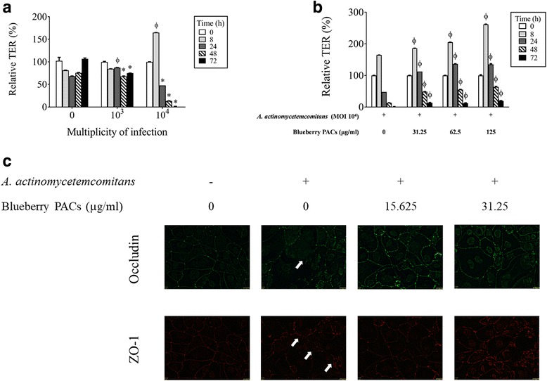
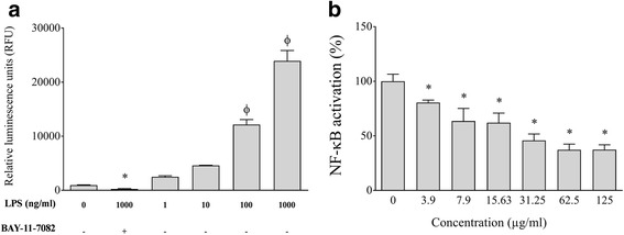
Methods: PACs were isolated from frozen highbush blueberries using solid-phase chromatography. A microplate dilution assay was performed to determine the effect of highbush blueberry PACs on A. actinomycetemcomitans growth as well as biofilm formation stained with crystal violet. Tight junction integrity of oral keratinocytes was assessed by measuring the transepithelial electrical resistance (TER), while macrophage viability was determined with a colorimetric MTT assay. Pro-inflammatory cytokine and MMP secretion by A. actinomycetemcomitans-stimulated macrophages was quantified by ELISA. The U937-3xκB-LUC monocyte cell line transfected with a luciferase reporter gene was used to monitor NF-κB activation.

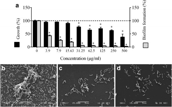
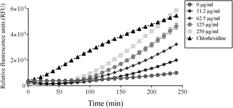
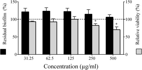
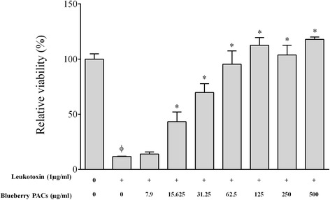
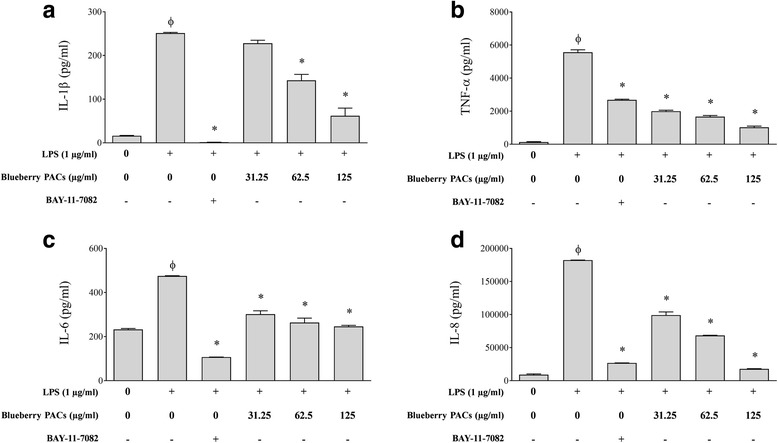
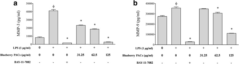
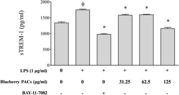
Results: Highbush blueberry PACs reduced the growth of A. actinomycetemcomitans and prevented biofilm formation at sub-inhibitory concentrations. The treatment of pre-formed biofilms with the PACs resulted in a loss of bacterial viability. The antibacterial activity of the PACs appeared to involve damage to the bacterial cell membrane. The PACs protected the oral keratinocytes barrier integrity from damage caused by A. actinomycetemcomitans. The PACs also protected macrophages from the deleterious effect of leukotoxin Ltx-A and dose-dependently inhibited the secretion of pro-inflammatory cytokines (IL-1β, IL-6, CXCL8, TNF-α), matrix metalloproteinases (MMP-3, MMP-9), and sTREM-1 by A. actinomycetemcomitans-treated macrophages. The PACs also inhibited the activation of the NF-κB signaling pathway.

Conclusion: The antibacterial and anti-inflammatory properties of highbush blueberry PACs as well as their ability to protect the oral keratinocyte barrier and neutralize leukotoxin activity suggest that they may be promising candidates as novel therapeutic agents.

Keywords: Aggregatibacter actinomycetemcomitans; Blueberry; Cytokine; Keratinocyte; Leukotoxin; Macrophages; Matrix metalloproteinase; Periodontal disease; Proanthocyanidins; Tight junction.


Conflict of interest statement

Ethics approval and consent to participate

Not applicable

Consent for publication

Not applicable

Competing interests

The authors declare that they have no competing interests.

Publisher’s Note

Springer Nature remains neutral with regard to jurisdictional claims in published maps and institutional affiliations.

Figures

Fig. 1
Fig. 1
Effect of highbush blueberry PACs on A. actinomycetemcomitans growth and biofilm formation (panel a). ∗: significantly different (p < 0.01) from the control (no PACs). Scanning electron micrographs of A. actinomycetemcomitans biofilms formed in the absence (panel b) and presence of 31.25 μg/ml (panel c) and 62.5 μg/ml of highbush blueberry PACs (panel d)
Fig. 2
Fig. 2
Time-course release of calcein-AM by A. actinomycetemcomitans cells treated with various concentrations of highbush blueberry PACs. Chlorhexidine (200 μg/ml) was used as a positive control. A significant (p < 0.01) release of calcein-AM was observed with PACs at 62.5, 125, and 250 μg/ml following a treatment of bacteria for 120 min. To obtain the same significance, a treatment of 160 min was necessary when PACs were used at 31.25 μg/ml
Fig. 3
Fig. 3
Effect of highbush blueberry PACs on A. actinomycetemcomitans biofilm desorption and viability. ∗: significantly different (p < 0.01) from the control (no PACs; horizontal line)
Fig. 4
Fig. 4
Effect of A. actinomycetemcomitans in the absence and presence of highbush blueberry PACs on the integrity of the oral keratinocyte tight junction. Panel a: Effects of time and the number of A. actinomycetemcomitans cells on the transepithelial electrical resistance (TER) of oral keratinocytes. Panel b: Inhibitory effect of highbush blueberry PACs on the A. actinomycetemcomitans-induced decrease in the TER of oral keratinocytes. Panel c: Immunofluorescence staining of tight junction proteins occludin and zonula occludens-1 (ZO-1) of oral keratinocytes infected (24 h) by A. actinomycetemcomitans in the absence and presence of blueberry PACs. A 100% value was assigned to the TER at time 0. Results are expressed as the mean ± SD of triplicate assays. Φ: significant increase (p < 0.001) compared with unstimulated control cells (a) or with A. actinomycetemcomitans-stimulated cells not treated with highbush blueberry PACs (B). ∗: significant decrease (p < 0.001) compared with unstimulated control cells
Fig. 5
Fig. 5
Effect of highbush blueberry PACs on A. actinomycetemcomitans leukotoxin (LtxA) activity on macrophage-like cells. Φ: significant loss of cell viability (p < 0.001) compared to control cells not treated with LtxA. *: significant increase of cell viability (p < 0.001) compared to cells treated with LtxA
Fig. 6
Fig. 6
Effect of highbush blueberry PACs on the secretion of a IL-1β, b TNF-α, c IL-6, and d CXCL8 by macrophages stimulated with A. actinomycetemcomitans LPS. The commercial inhibitor BAY-11-7082 (25 μM) was used as a positive inhibitory control. Results are expressed as the means ± SD of triplicate assays from three independent experiments. Φ: significant increase (p < 0.001) compared to unstimulated control cells. *: significant decrease (p < 0.001) compared to LPS-stimulated cells not treated with highbush blueberry PACs
Fig. 7
Fig. 7
Effect of highbush blueberry PACs on the secretion of a MMP-3 and b MMP-9 by macrophages stimulated with A. actinomycetemcomitans LPS. The commercial inhibitor BAY-11-7082 (25 μM) was used as a positive inhibitory control. Results are expressed as the means ± SD of triplicate assays from three independent experiments. Φ: significant increase (p < 0.001) compared to unstimulated control cells. *: significant decrease (p < 0.001) compared to LPS-stimulated cells not treated with highbush blueberry PACs
Fig. 8
Fig. 8
Effect of highbush blueberry PACs on the secretion/shedding of sTREM-1 by macrophages stimulated with A. actinomycetemcomitans LPS. The commercial inhibitor BAY-11-7082 was used as a positive inhibitory control. Results are expressed as the means ± SD of triplicate assays from three independent experiments. Φ: significant increase (p < 0.001) compared to non-stimulated control cells. *: significant decrease (p < 0.001) compared to LPS-stimulated cells not treated with highbush blueberry PACs
Fig. 9
Fig. 9
Effect of A. actinomycetemcomitans LPS in the absence and presence of highbush blueberry PACs on nuclear factor-κB (NF-κB) activation using the U937-3xκB cell model. Panel a Concentration effect of A. actinomycetemcomitans LPS on NF-κB activation. Panel b Inhibitory effect of highbush blueberry PACs on A. actinomycetemcomitans LPS-induced NF-κB activation. A value of 100% was assigned to the activation obtained with A. actinomycetemcomitans LPS in the absence of highbush blueberry PACs. BAY-11-7082 was used as a positive inhibitory control. Results are expressed as the mean ± SD of triplicate assays from two independent experiments. Φ: significant increase (p < 0.001) compared with nonstimulated control cells. ∗: significant decrease (p < 0.001) compared with LPS-stimulated cells not treated with highbush blueberry PACs

References

    1. Socransky SS, Haffajee AD. The bacterial etiology of destructive periodontal disease: current concepts. J Periodontol. 1992;63(4 Suppl):322–331. - PubMed
    1. Fine DH, Markowitz K, Furgang D, Fairlie K, Ferrandiz J, Nasri C, McKiernan M, Gunsolley J. Aggregatibacter actinomycetemcomitans and its relationship to initiation of localized aggressive periodontitis: longitudinal cohort study of initially healthy adolescents. J Clin Microbiol. 2007;45(12):3859–3869. - PMC - PubMed
    1. Zambon JJ, Christersson LA, Slots J. Actinobacillus actinomycetemcomitans in human periodontal disease. Prevalence in patient groups and distribution of biotypes and serotypes within families. J Periodontol. 1983;54(12):707–711. - PubMed
    1. van Winkelhoff AJ, Slots J. Actinobacillus actinomycetemcomitans and Porphyromonas gingivalis in nonoral infections. Periodontol 2000. 1999;20:122–135. - PubMed
    1. Henderson B, Ward JM, Ready D. Aggregatibacter (Actinobacillus) actinomycetemcomitans: a triple A* periodontopathogen? Periodontol 2000. 2010;54(1):78–105. - PubMed

MeSH terms

LinkOut - more resources