Review
. 2018 Mar;29(3):759-774.
doi: 10.1681/ASN.2017090958. Epub 2018 Jan 10.

Differentiating Primary, Genetic, and Secondary FSGS in Adults: A Clinicopathologic Approach

Affiliations
Review

Differentiating Primary, Genetic, and Secondary FSGS in Adults: A Clinicopathologic Approach

An S De Vriese et al. J Am Soc Nephrol. 2018 Mar.

Abstract

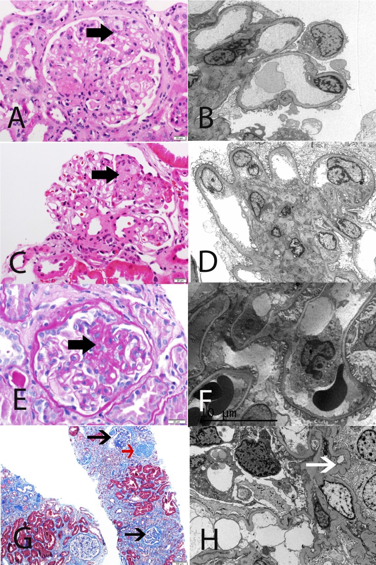
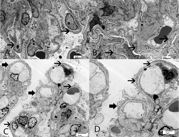
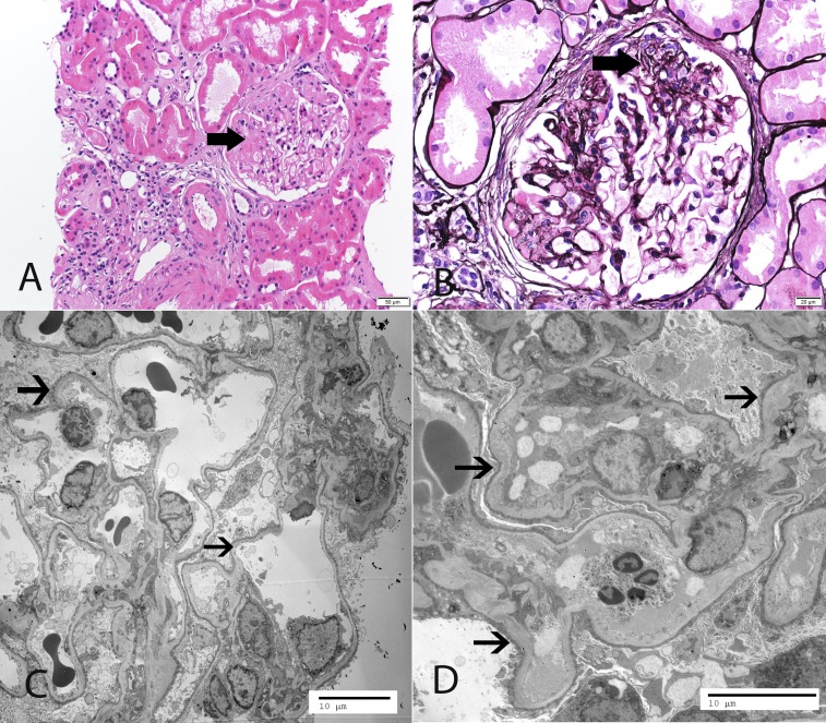
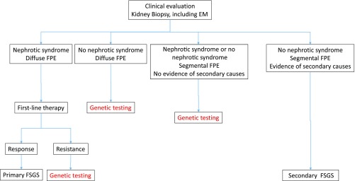
FSGS describes a renal histologic lesion with diverse causes and pathogenicities that are linked by podocyte injury and depletion. Subclasses of FSGS include primary, genetic, and secondary forms, the latter comprising maladaptive, viral, and drug-induced FSGS. Despite sharing certain clinical and histologic features, these subclasses differ noticeably in management and prognosis. Without an accepted nongenetic biomarker that discriminates among these FSGS types, classification of patients is often challenging. This review summarizes the clinical and histologic features, including the onset and severity of proteinuria as well as the presence of nephrotic syndrome, that may aid in identifying the specific FSGS subtype. The FSGS lesion is characterized by segmental sclerosis and must be differentiated from nonspecific focal global glomerulosclerosis. No light microscopic features are pathognomonic for a particular FSGS subcategory. The characteristics of podocyte foot process effacement on electron microscopy, while helpful in discriminating between primary and maladaptive FSGS, may be of little utility in detecting genetic forms of FSGS. When FSGS cannot be classified by clinicopathologic assessment, genetic analysis should be offered. Next generation DNA sequencing enables cost-effective screening of multiple genes simultaneously, but determining the pathogenicity of a detected genetic variant may be challenging. A more systematic evaluation of patients, as suggested herein, will likely improve therapeutic outcomes and the design of future trials in FSGS.

Keywords: FSGS; Primary; Secondary; genetic renal disease; nephrotic syndrome.


Figures

Figure 1.
Figure 1.
Representative LM and EM findings in FSGS and FGGS. (A and B) Primary FSGS in a 61-year-old white man with serum creatinine 1.5 mg/dl, proteinuria 14.2 g/24 h, and serum albumin 2.5 mg/dl. LM shows segmental sclerosis, and EM highlights diffuse FPE. (C and D) Maladaptive FSGS in a 53-year-old white man with serum creatinine 1.9 mg/dl, proteinuria 5.8 g/24 h, and serum albumin 4.1 mg/dl. LM shows segmental sclerosis, and EM reveals well preserved foot processes. (E and F) Genetic FSGS in a 39-year-old white man with familial history of PLCE1 mutation, serum creatinine 1.3 mg/dl, proteinuria 2.0 g/24 h, and serum albumin 4 g/dl. LM shows segmental sclerosis, and EM features only minimal FPE. (G and H) FGGS in a 38-year-old black man with a long history of uncontrolled hypertension, serum creatinine 5.1 mg/dl, proteinuria 2.8 g/24 h, and serum albumin 4.3 g/dl. LM shows ischemic changes, as well as a globally sclerosed glomerulus (red arrow), and EM shows relatively well preserved foot processes. Note ischemic capillary loops (white arrow). (A, C, E) Thick black arrow points to segmental sclerosis, and (G) thin black arrow points to ischemic glomeruli. (A and C) Hematoxylin and eosin. (E) Periodic acid–Schiff. (G) Masson trichrome stain. Original magnification, ×40 in A, C, and E; ×2500 in B; ×3000 in D; ×3500 in F; ×10 in G; ×5000 in H.
Figure 2.
Figure 2.
EM findings in two siblings who are both compound heterozygotes for the NPHS2 mutation and the R286fs pathogenic variant along with a non-neutral polymorphism R229Q. (A and B) The 63-year-old brother with serum creatinine 1.1 mg/dl, proteinuria 6.6 g/24 h, and serum albumin 3.4 g/dl. EM shows diffuse FPE. (C and D) The 47-year-old sister with serum creatinine 0.9 mg/dl, proteinuria 2.5 g/24 h, and serum albumin 4 g/dl. EM shows segmental FPE. Thick black arrow points to preserved foot processes, and thin black arrow points to effaced foot processes. Original magnification, ×5000 in A and B; ×2900 in C; ×4800 in D.
Figure 3.
Figure 3.
Representative LM and EM findings. A 46-year-old white woman, in whom the initial biopsy at age 18 years old was reported as minimal change disease, presented with serum creatinine 1.5 mg/dl, proteinuria 5.8 g/24 h, and serum albumin 3.4 g/dl. The patient was resistant to corticosteroids and calcineurin inhibitors. Mutation analysis with a next generation sequencing panel showed two mutations in NPHS2 and a heterozygous variation in WDR73, which is known to cause the Galloway–Mowat syndrome. (A and B) LM shows segmental sclerosis (thick arrows point to segmental sclerosis). (C and D) EM reveals diffuse FPE (thin arrows point to effaced foot processes). (A) Hematoxylin and eosin. (B) Silver methenamine stain. Original magnification, ×20 in A; ×40 in B; ×1900 in C; ×2900 in D.
Figure 4.
Figure 4.
Opinion-based approach to genetic testing in adult-onset FSGS. Note that viral- and drug-associated forms of FSGS are usually excluded by clinical and serologic evaluation.

References

    1. D’Agati VD, Fogo AB, Bruijn JA, Jennette JC: Pathologic classification of focal segmental glomerulosclerosis: A working proposal. Am J Kidney Dis 43: 368–382, 2004 - PubMed
    1. Kachurina N, Chung CF, Benderoff E, Babayeva S, Bitzan M, Goodyer P, Kitzler T, Matar D, Cybulsky AV, Alachkar N, Torban E: Novel unbiased assay for circulating podocyte-toxic factors associated with recurrent focal segmental glomerulosclerosis. Am J Physiol Renal Physiol 310: F1148–F1156, 2016 - PubMed
    1. Allard L, Kwon T, Krid S, Bacchetta J, Garnier A, Novo R, Deschenes G, Salomon R, Roussey G, Allain-Launay E: Treatment by immunoadsorption for recurrent focal segmental glomerulosclerosis after paediatric kidney transplantation: A multicentre French cohort [published online ahead of print July 28, 2017]. Nephrol Dial Transplant 10.1093/ndt/gfx214 - DOI - PubMed
    1. D’Agati VD: Podocyte growing pains in adaptive FSGS. J Am Soc Nephrol 28: 2825–2827, 2017 - PMC - PubMed
    1. Chandra P, Kopp JB: Viruses and collapsing glomerulopathy: A brief critical review. Clin Kidney J 6: 1–5, 2013 - PMC - PubMed

MeSH terms