Comparative Study
. 2018 Feb 23;293(8):2815-2828.
doi: 10.1074/jbc.RA117.000656. Epub 2018 Jan 10.

Engineered synthetic antibodies as probes to quantify the energetic contributions of ligand binding to conformational changes in proteins

Affiliations
Comparative Study

Engineered synthetic antibodies as probes to quantify the energetic contributions of ligand binding to conformational changes in proteins

Somnath Mukherjee et al. J Biol Chem. .

Abstract

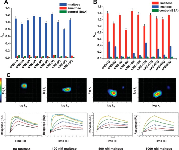
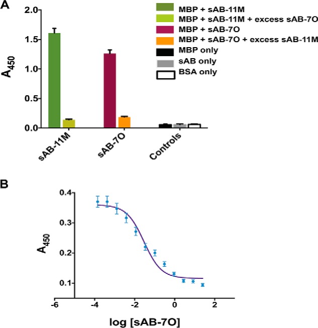
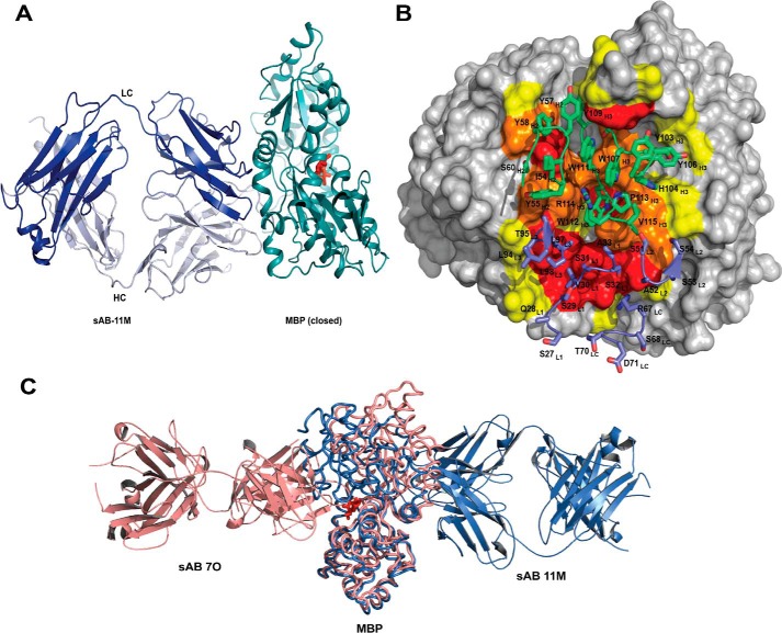
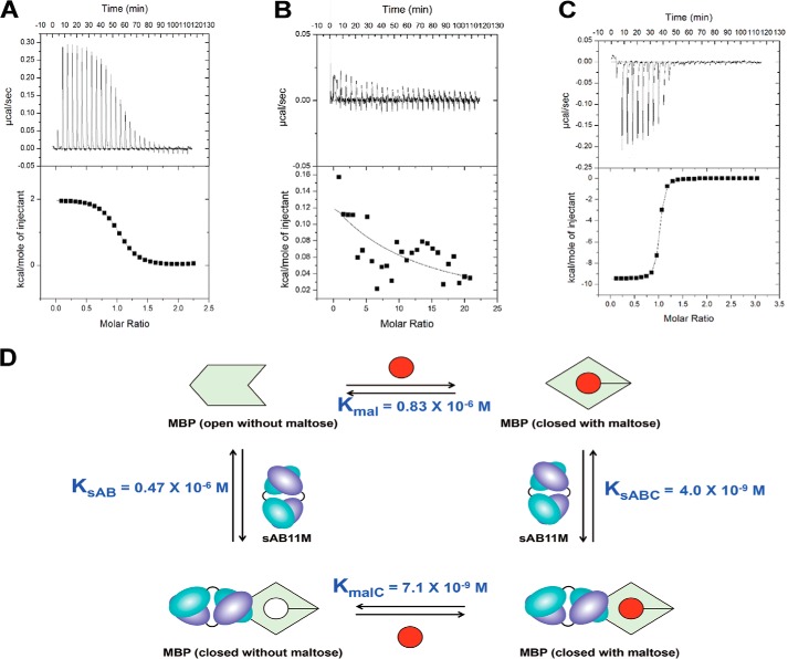
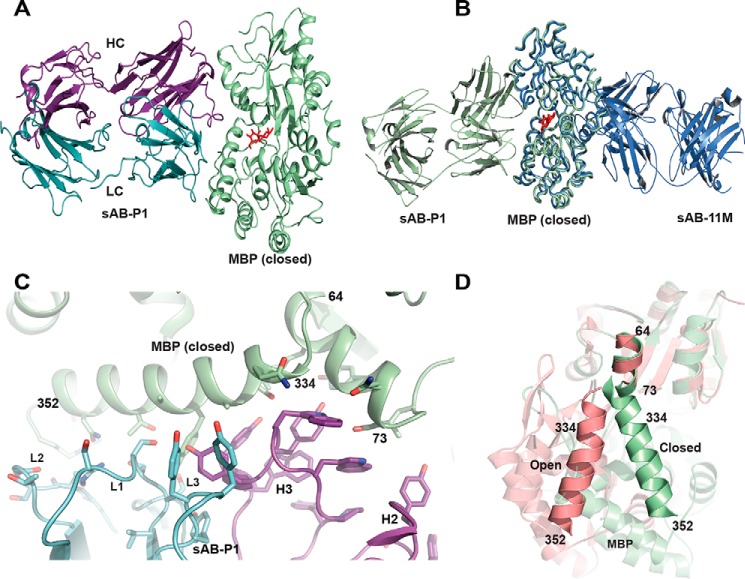
Conformational changes in proteins due to ligand binding are ubiquitous in biological processes and are integral to many biological systems. However, it is often challenging to link ligand-induced conformational changes to a resulting biological function because it is difficult to distinguish between the energetic components associated with ligand binding and those due to structural rearrangements. Here, we used a unique approach exploiting conformation-specific and regio-specific synthetic antibodies (sABs) to probe the energetic contributions of ligand binding to conformation changes. Using maltose-binding protein (MBP) as a model system, customized phage-display selections were performed to generate sABs that stabilize MBP in different conformational states, modulating ligand-binding affinity in competitive, allosteric, or peristeric manners. We determined that the binding of a closed conformation-specific sAB (sAB-11M) to MBP in the absence of maltose is entropically driven, providing new insight into designing antibody-stabilized protein interactions. Crystal structures of sABs bound to MBP, together with biophysical data, delineate the basis of free energy differences between different conformational states and confirm the use of the sABs as energy probes for dissecting enthalpic and entropic contributions to conformational transitions. Our work provides a foundation for investigating the energetic contributions of distinct conformational dynamics to specific biological outputs. We anticipate that our approach also may be valuable for analyzing the energy landscapes of regulatory proteins controlling biological responses to environmental changes.

Keywords: allostery; antibody engineering; conformation-specific synthetic antibodies; crystal structure; energy probes; epitope mapping; phage display; thermodynamics.


Conflict of interest statement

The authors declare that they have no conflicts of interest with the contents of this article

Figures

Figure 1.
Figure 1.
Characterization of conformation-specific sABs. Phage ELISA shows that sABs generated against the open form of MBP in the absence of maltose do not bind to the closed maltose-bound form of MBP (A) and sABs generated against the maltose-bound, closed form of MBP do not bind or bind with very reduced affinity to the open form of MBP (B). C, kinetics of sAB-7O binding to MBP at different maltose concentration. The kinetics is on-rate–dependent, and affinity decreases steadily with increasing maltose concentration. Interaction Map shows a homogeneous 1:1 interaction. Change in relative position of the heat map on the y axis (log ka) is clear due to a decrease in the on-rate with maltose concentration, whereas the position on the x axis (log kd) remains constant due to invariance in the off-rate. Error bars, S.E.
Figure 2.
Figure 2.
Closed and open conformation–specific sABs are competitive. A, single-point ELISA shows that the binding of sAB-7O and sAB-11M are exclusive. Neither of them binds in the presence of an excess of the other. B, competitive ELISA showing sAB-7O displacing His6-sAB-11M from MBP saturated with His6-sAB-11M. In both A and B, the sAB of interest was His-tagged, and the ELISA signal (A450) was monitored using anti-His antibody. Error bars, S.D.
Figure 3.
Figure 3.
Structures of open-specific sAB-7O·MBP complex. A, sAB-7O binds to MBP in open conformation by the light chain CDRs (L1 and L3) and heavy chain CDRs (H1, H2, and H3). B, open-book view of the interface between sAB-7O and MBP. Residues in the interface are colored according to their percentage of reduction in accessible surface area upon complex formation (yellow, 10–49%; orange, 50–70%; red, >70%). C, CDR-H3 residues of sAB-7O (green) interact with the residues in the maltose-binding pocket of MBP (salmon). D, residues of MBP (salmon) involved in maltose binding. Selected hydrogen-bonding interactions with maltose (ball and stick) are highlighted. Most of the maltose-binding residues in MBP are involved in interaction with CDR-H3 residues of sAB-7O as seen in C. Sugar rings and hydroxyl groups of maltose are mimicked by the aromatic ring of tyrosine and backbone carbonyls in CDR-H3 residues, resulting in strong bias of the maltose binding pocket as the sole immunodominant epitope.
Figure 4.
Figure 4.
Structures of closed-specific sAB-11M·MBP complex. A, sAB-11M binds to the hinge region in the maltose (red sticks)-bound conformation of MBP on the side opposite to the maltose-binding pocket. B, detailed picture of sAB-11M residues (shown in sticks) interacting with MBP. CDR-L1, -L2, and -L3 and LC scaffold residues are colored marine, and CDR-H1, -H2, and -H3 are colored green. MBP is color-coded as in Fig. 3B. C, superposition of structures of sAB-7O-MBP structure (salmon) with sAB-11M-MBP (slate) shows that sAB-7O and sAB-11M bind on opposite sides of MBP in open and closed states, respectively.
Figure 5.
Figure 5.
Thermodynamics of maltose and sABs binding to MBP. Shown are ITC titration curves of maltose binding to MBP (A), MBP with a 5-fold excess of sAB-7O (B), and MBP with 5-fold excess of sAB-11M (C). D, thermodynamic cycle of maltose binding to MBP with and without sAB-11M.
Figure 6.
Figure 6.
Structural features of sAB-P1·MBP complex. A, sAB-P1 binds to the closed conformation of MBP interacting across the face of the binding pocket with the maltose trapped inside. B, superposition of MBP bound to sAB-P1 (green) with that of sAB-11M (slate) shows that sAB-P1 binds to the peristeric site of MBP in the closed form opposite to sAB-11M. C, interactions of the HC (magenta) and LC (blue) CDRs of sAB-P1 with the two helices (64–73 and 334–352) of MBP (green) at the peristeric interface. D, the relative orientations of the helices comprising residues 64–73 and 341–350 are different in open (salmon) and closed (green) forms of MBP. These helices in the closed conformation interact extensively with sAB-P1 (C), thereby imparting conformational specificity.

Comment in

References

    1. Aaronson D. S., and Horvath C. M. (2002) A road map for those who know JAK-STAT. Science 296, 1653–1655 10.1126/science.1071545 - DOI - PubMed
    1. Oehler S., Eismann E. R., Krämer H., and Müller-Hill B. (1990) The three operators of the lac operon cooperate in repression. EMBO J. 9, 973–979 - PMC - PubMed
    1. Yonetani T., Park S. I., Tsuneshige A., Imai K., and Kanaori K. (2002) Global allostery model of hemoglobin: modulation of O2 affinity, cooperativity, and Bohr effect by heterotropic allosteric effectors. J. Biol. Chem. 277, 34508–34520 10.1074/jbc.M203135200 - DOI - PubMed
    1. Sharff A. J., Rodseth L. E., Spurlino J. C., and Quiocho F. A. (1992) Crystallographic evidence of a large ligand-induced hinge-twist motion between the two domains of the maltodextrin binding protein involved in active transport and chemotaxis. Biochemistry 31, 10657–10663 10.1021/bi00159a003 - DOI - PubMed
    1. Millet O., Hudson R. P., and Kay L. E. (2003) The energetic cost of domain reorientation in maltose-binding protein as studied by NMR and fluorescence spectroscopy. Proc. Natl. Acad. Sci. U.S.A. 100, 12700–12705 10.1073/pnas.2134311100 - DOI - PMC - PubMed

Publication types

MeSH terms