Review
. 2018 Jan 10;131(1):jcs203760.
doi: 10.1242/jcs.203760.

Reconsidering an active role for G-actin in cytoskeletal regulation

Affiliations
Review

Reconsidering an active role for G-actin in cytoskeletal regulation

Kristen Skruber et al. J Cell Sci. .

Abstract

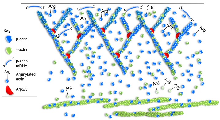
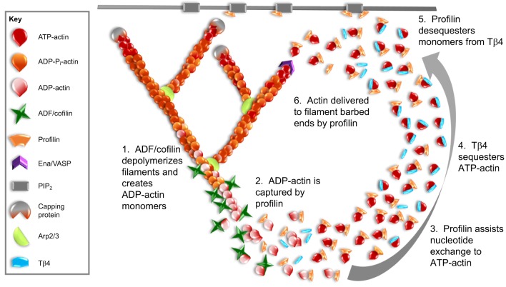
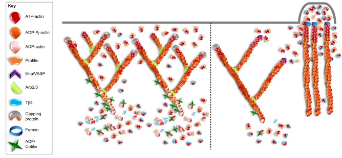
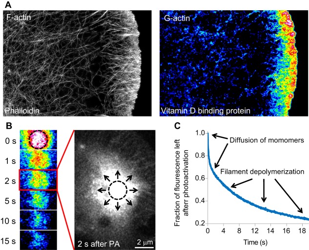
Globular (G)-actin, the actin monomer, assembles into polarized filaments that form networks that can provide structural support, generate force and organize the cell. Many of these structures are highly dynamic and to maintain them, the cell relies on a large reserve of monomers. Classically, the G-actin pool has been thought of as homogenous. However, recent work has shown that actin monomers can exist in distinct groups that can be targeted to specific networks, where they drive and modify filament assembly in ways that can have profound effects on cellular behavior. This Review focuses on the potential factors that could create functionally distinct pools of actin monomers in the cell, including differences between the actin isoforms and the regulation of G-actin by monomer binding proteins, such as profilin and thymosin β4. Owing to difficulties in studying and visualizing G-actin, our knowledge over the precise role that specific actin monomer pools play in regulating cellular actin dynamics remains incomplete. Here, we discuss some of these unanswered questions and also provide a summary of the methodologies currently available for the imaging of G-actin.

Keywords: G-actin; Profilin; Thymosin β 4; β-actin; γ-actin.


Conflict of interest statement

Competing interestsThe authors declare no competing or financial interests.

Figures

Fig. 1.
Fig. 1.
Incorporation of β-actin and γ-actin into distinct actin networks. Although they can overlap, β-actin and γ-actin isoforms have been shown to exhibit different cellular localizations. Illustrated here is a scenario where β-actin is enriched in the lamellipodia and γ-actin in actin arcs and/or stress fiber structures. β-actin-encoding mRNA is also localized and locally translated at the leading edge. Arginylation of β-actin (Arg) promotes localization and assembly in lamellipodia but targets γ-actin for degradation, as depicted by the pale color.
Fig. 2.
Fig. 2.
Profilin and Tβ4 regulate the monomer pool. This figure presents a model for how profilin and Tβ4 may work together to convert newly released actin monomers into a polymerization-competent pool that is then directed to re-polymerize into new filaments. After depolymerization of filaments by disassembly factors such as ADF/cofilin (1), profilin binds to the newly released monomers (2) due to its higher affinity for ADP-actin. Profilin induces nucleotide exchange (3), and the majority of monomers are transferred to Tβ4 (4), which has a 50-fold higher affinity for ATP-actin over ADP-actin and is present in the cell at higher concentrations than profilin. Tβ4 holds the monomers in a polymerization-competent pool. Owing to high rates of exchange, profilin de-sequesters monomers from Tβ4 (5) and delivers G-actin to barbed-end polymerases such as Ena/VASP (6). Profilin can also localize to the plasma membrane through an interaction with phosphatidylinositol 4,5-bisphosphate (PIP2). A profilin–Tβ4–G-actin ternary complex may assist in the transfer of G-actin from Tβ4 to profilin by providing an intermediate state, where the actin monomer does not dissociate from either protein, thereby preventing its spontaneous assembly into filaments.
Fig. 3.
Fig. 3.
G-actin regulated assembly of actin networks. A scenario where profilin and Tβ4 regulate the assembly of specific actin networks. On the left, there is increased ADF/cofilin and capping protein activity; this leads to increased disassembly and barbed-end capping, thus resulting in an increased monomer pool. This creates a more densely branched Arp2/3-mediated actin network because nucleation, rather than elongation, becomes favored. Here, the role of profilin is predominantly to recycle the monomers back into a polymerization-competent pool. On the right, ADF/cofilin and capping protein activity is decreased, resulting in a smaller monomer pool. Consequently, competition for a limited supply of monomers enhances filament elongation by barbed-end polymerases, such as Ena/VASP and formins, and reduces Arp2/3-mediated branching due to a bias formed through profilin-bound actin. Additionally, Tβ4 prevents cytosolic actin monomers from polymerizing at barbed ends of Arp2/3-mediated filaments throughout the lamellipodia (right-hand side of this panel) and selectively releases G-actin near the plasma membrane, where it undergoes formin-mediated polymerization.
Fig. 4.
Fig. 4.
Methods for visualizing G-actin. (A) Actin monomers can be visualized in fixed cells with monomer-specific probes such as vitamin D-binding protein. Here, structured illumination super-resolution images show the same cell stained with both phalloidin, to visualize F-actin, and vitamin D-binding protein, to visualize actin monomers. The vitamin D-binding protein image is pseudocolored to emphasize changes in fluorescence, with warmer colors representing increased fluorescence. Actin monomers can be seen localized to the leading edge. (B) G-actin can be visualized in live cells by measuring rapid changes in fluorescently labeled actin. Here, a pulse-chase experiment is performed using actin labeled with photoactivatable GFP. After photoactivation (PA, occurring in the region marked by the dotted circle), the actin is highlighted and can be followed over time. The G-actin rapidly diffuses away from the PA region in seconds (represented by the arrows in the magnified image), which is emphasized by the middle panel showing a larger image of the cell two seconds after actin was photoactivated. (C) A representative graph from this type of experiment shown in B, highlighting which parts of the fluorescence decay curve are due to diffusion of monomers away from the photoactivated region and which parts arise from the disassembly of F-actin that occurs on a slower time scale. Images in A are similar to those published in Lee et al., 2013; images in B are similar to those published in Kapustina et al., 2016.

References

    1. Agbulut O., Huet A., Niederländer N., Puceat M., Menasché P. and Coirault C. (2007). Green fluorescent protein impairs actin-myosin interactions by binding to the actin-binding site of myosin. J. Biol. Chem. 282, 10465-10471. 10.1074/jbc.M610418200 - DOI - PubMed
    1. Aizawa H., Sameshima M. and Yahara I. (1997). A green fluorescent protein-actin fusion protein dominantly inhibits cytokinesis, cell spreading, and locomotion in Dictyostelium. Cell Struct. Funct. 22, 335-345. 10.1247/csf.22.335 - DOI - PubMed
    1. Akin O. and Mullins R. D. (2008). Capping protein increases the rate of actin-based motility by promoting filament nucleation by the Arp2/3 complex. Cell 133, 841-851. 10.1016/j.cell.2008.04.011 - DOI - PMC - PubMed
    1. Andrianantoandro E. and Pollard T. D. (2006). Mechanism of actin filament turnover by severing and nucleation at different concentrations of ADF/cofilin. Mol. Cell 24, 13-23. 10.1016/j.molcel.2006.08.006 - DOI - PubMed
    1. Baranwal S., Naydenov N. G., Harris G., Dugina V., Morgan K. G., Chaponnier C. and Ivanov A. I. (2012). Nonredundant roles of cytoplasmic beta- and gamma-actin isoforms in regulation of epithelial apical junctions. Mol. Biol. Cell 23, 3542-3553. 10.1091/mbc.E12-02-0162 - DOI - PMC - PubMed

Publication types

LinkOut - more resources