Review
. 2018 Jan;15(138):20170821.
doi: 10.1098/rsif.2017.0821.

Computational techniques for ECG analysis and interpretation in light of their contribution to medical advances

Affiliations
Review

Computational techniques for ECG analysis and interpretation in light of their contribution to medical advances

Aurore Lyon et al. J R Soc Interface. 2018 Jan.

Abstract

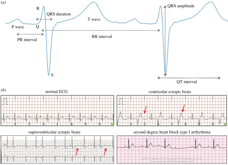
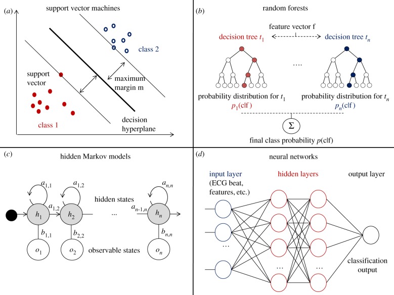
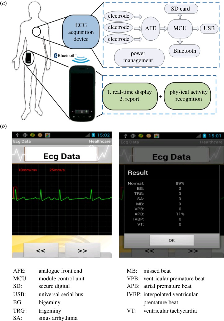
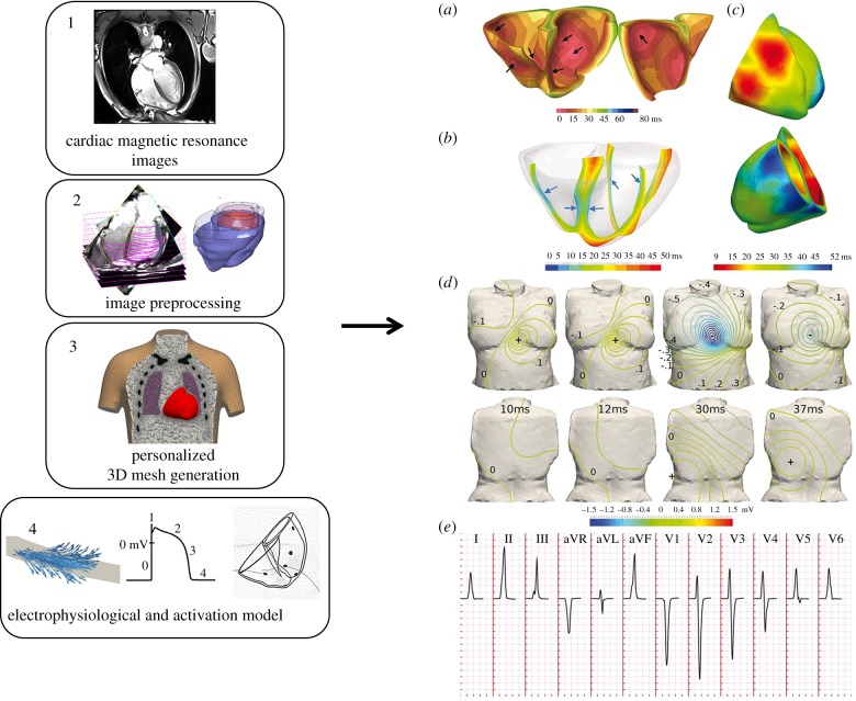
Widely developed for clinical screening, electrocardiogram (ECG) recordings capture the cardiac electrical activity from the body surface. ECG analysis can therefore be a crucial first step to help diagnose, understand and predict cardiovascular disorders responsible for 30% of deaths worldwide. Computational techniques, and more specifically machine learning techniques and computational modelling are powerful tools for classification, clustering and simulation, and they have recently been applied to address the analysis of medical data, especially ECG data. This review describes the computational methods in use for ECG analysis, with a focus on machine learning and 3D computer simulations, as well as their accuracy, clinical implications and contributions to medical advances. The first section focuses on heartbeat classification and the techniques developed to extract and classify abnormal from regular beats. The second section focuses on patient diagnosis from whole recordings, applied to different diseases. The third section presents real-time diagnosis and applications to wearable devices. The fourth section highlights the recent field of personalized ECG computer simulations and their interpretation. Finally, the discussion section outlines the challenges of ECG analysis and provides a critical assessment of the methods presented. The computational methods reported in this review are a strong asset for medical discoveries and their translation to the clinical world may lead to promising advances.

Keywords: classification; computer simulations; electrocardiogram; interpretation and analysis; machine learning.


Conflict of interest statement

We have no competing interests.

Figures

Figure 1.
Figure 1.
Example of data available for the analysis of ECG signals. (a) ECG waveforms (P, QRS and T waves) and standard features extracted from an ECG beat. The RR interval is measured as the peak-to-peak interval between two consecutive QRS complexes, the PR interval is defined as the duration from the beginning of the P wave to the beginning of the QRS complex, the QRS duration (or width) is the duration between the start and the end of the QRS complex, the QRS amplitude is defined as the absolute value of the difference between the minimum and the maximum of the QRS complex, and the QT interval is measured as the time between the beginning of the Q wave and the end of the T wave. (b) Examples of different ECG waveforms: normal ECG [50], ventricular ectopic beats [51], supraventricular ectopic beats [52] and second degree heart block arrhythmia [53]. (Online version in colour.)
Figure 2.
Figure 2.
Main machine learning methods used for ECG classification. (a) Support vector machine binary classification by maximization of the margin m. (b) Random forest classification using n decision trees. (c) Hidden Markov model with n states, transition matrix (ai,j) and emission matrix (bi,j). (d) Neural network with two hidden layers. (Online version in colour.)
Figure 3.
Figure 3.
Example of an ECG classification system on wearable device showing the methodology (a), and output (b). From Miao et al. [80]. Panel a details the ECG acquisition sensor implanted in the wearable device: the signal, recorded by electrodes, is amplified and filtered by the AFE module, converted to digital signal by the MCU module and recorded on the SD card, transmitted to the USB port or sent directly to the phone via Bluetooth. Panel b describes the output panel provided to the user: a screenshot of the ECG excerpt (left), and a summary of the normal and abnormal beats recorded (right).
Figure 4.
Figure 4.
Computational pipeline for ECG simulation from magnetic resonance images through 3D meshes and cellular electrophysiological models (left, from Zacur et al. [87]) and the obtained simulation of the cardiac electrical activity (right, from Cardone-Noott et al. [13]). Personalized cardiac magnetic resonance images (1) are segmented and preprocessed (2). From this information, surface and volumetric meshes are generated (3) and an electrophysiological model defining the electrical activity in the cells is implemented (4). The obtained simulated electrical conduction (right panel) can then be investigated.

References

    1. Institute of Medicine (US) Committee on Preventing the Global Epidemic of Cardiovascular Disease: Meeting the Challenges in Developing Countries. 2010. Epidemiology of cardiovascular disease. In Promoting cardiovascular health in the developing world: a critical challenge to achieve global health (eds Fuster V, Kelly BB). Washington, DC: National Academies Press; See https://www.ncbi.nlm.nih.gov/books/NBK45688/ - PubMed
    1. Obermeyer Z, Emanuel EJ. 2016. Predicting the future—big data, machine learning, and clinical medicine. N. Engl. J. Med. 375, 1216–1219. (10.1056/NEJMp1606181) - DOI - PMC - PubMed
    1. Sörnmo L, Laguna P. 2005. Bioelectrical signal processing in cardiac and neurological applications (ed. LS Laguna). Burlington: Academic Press; See http://www.sciencedirect.com/science/article/pii/B9780124375529500015 (accessed 10 October 2014)
    1. Llamedo M, Martinez JP. 2011. Heartbeat classification using feature selection driven by database generalization criteria. IEEE Trans. Biomed. Eng. 58, 616–625. (10.1109/TBME.2010.2068048) - DOI - PubMed
    1. de Chazal P, Reilly RB. 2006. A patient-adapting heartbeat classifier using ECG morphology and heartbeat interval features. IEEE Trans. Biomed. Eng. 53, 2535–2543. (10.1109/TBME.2006.883802) - DOI - PubMed

Publication types