. 2018 Mar 14;92(7):e01654-17.
doi: 10.1128/JVI.01654-17. Print 2018 Apr 1.

Herpes Simplex Virus 1 Mutant with Point Mutations in UL39 Is Impaired for Acute Viral Replication in Mice, Establishment of Latency, and Explant-Induced Reactivation

Affiliations

Herpes Simplex Virus 1 Mutant with Point Mutations in UL39 Is Impaired for Acute Viral Replication in Mice, Establishment of Latency, and Explant-Induced Reactivation

Heba H Mostafa et al. J Virol. .

Abstract

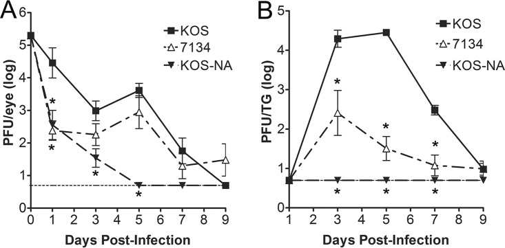
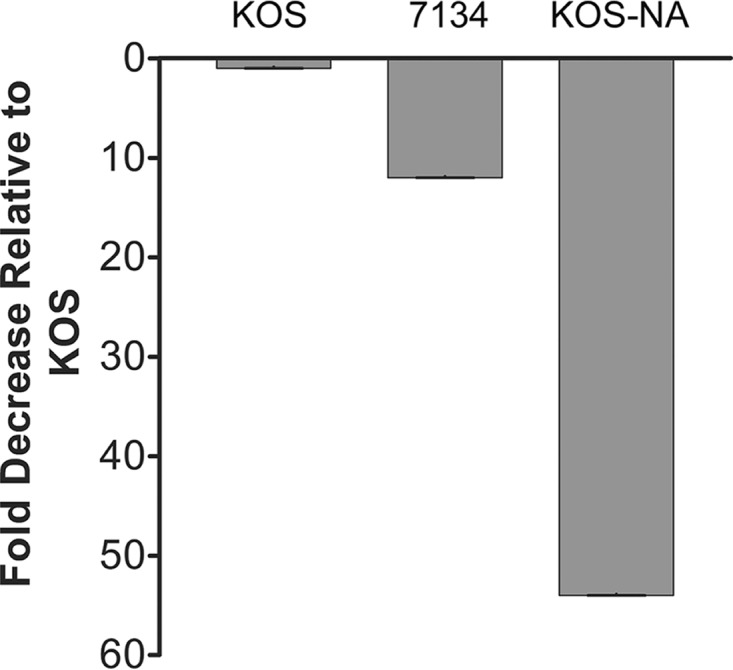
In the process of generating herpes simplex virus 1 (HSV-1) mutations in the viral regulatory gene encoding infected cell protein 0 (ICP0), we isolated a viral mutant, termed KOS-NA, that was severely impaired for acute replication in the eyes and trigeminal ganglia (TG) of mice, defective in establishing a latent infection, and reactivated poorly from explanted TG. To identify the secondary mutation(s) responsible for the impaired phenotypes of this mutant, we sequenced the KOS-NA genome and noted that it contained two nonsynonymous mutations in UL39, which encodes the large subunit of ribonucleotide reductase, ICP6. These mutations resulted in lysine-to-proline (residue 393) and arginine-to-histidine (residue 950) substitutions in ICP6. To determine whether alteration of these amino acids was responsible for the KOS-NA phenotypes in vivo, we recombined the wild-type UL39 gene into the KOS-NA genome and rescued its acute replication phenotypes in mice. To further establish the role of UL39 in KOS-NA's decreased pathogenicity, the UL39 mutations were recombined into HSV-1 (generating UL39mut), and this mutant virus showed reduced ocular and TG replication in mice comparable to that of KOS-NA. Interestingly, ICP6 protein levels were reduced in KOS-NA-infected cells relative to the wild-type protein. Moreover, we observed that KOS-NA does not counteract caspase 8-induced apoptosis, unlike wild-type strain KOS. Based on alignment studies with other HSV-1 ICP6 homologs, our data suggest that amino acid 950 of ICP6 likely plays an important role in ICP6 accumulation and inhibition of apoptosis, consequently impairing HSV-1 pathogenesis in a mouse model of HSV-1 infection.IMPORTANCE HSV-1 is a major human pathogen that infects ∼80% of the human population and can be life threatening to infected neonates or immunocompromised individuals. Effective therapies for treatment of recurrent HSV-1 infections are limited, which emphasizes a critical need to understand in greater detail the events that modulate HSV-1 replication and pathogenesis. In the current study, we identified a neuroattenuated HSV-1 mutant (i.e., KOS-NA) that contains novel mutations in the UL39 gene, which codes for the large subunit of ribonucleotide reductase (also known as ICP6). This mutant form of ICP6 was responsible for the attenuation of KOS-NA in vivo and resulted in diminished ICP6 protein levels and antiapoptotic effect. Thus, we have determined that subtle alteration of the UL39 gene regulates expression and functions of ICP6 and severely impacts HSV-1 pathogenesis, potentially making KOS-NA a promising vaccine candidate against HSV-1.

Keywords: HSV-1; ICP6; UL39; antiapoptosis; pathogenesis; ribonucleotide reductase; viral mutant.


Figures

FIG 1
FIG 1
Acute replication of KOS-NA in mice. Groups of mice were infected with 2 × 10⁵ PFU per eye. Tear film was collected on days 1, 3, 5, 7, and 9 postinfection, and TG were collected at the same time points from cohorts. Infectious virus in swab samples and homogenized TG was quantified by plaque assay. (A) Acute corneal replication of KOS-NA. (B) Acute replication of KOS-NA in TG. Results are geometric means ± SEMs (n = 6 samples per group per time point). The horizontal dotted lines represent the lower limit of detection. *, Student's t test, P < 0.05 compared with KOS. A portion of the control data was previously presented in reference .
FIG 2
FIG 2
Viral genome loads in latent TG. Mice were infected with 2 × 10⁵ PFU per eye, and TG were collected 28 to 30 days postinfection. DNA was extracted from latent TG, and the amount of HSV-1 DNA present was quantified by real-time PCR (n = 4 to 10 TG per group). Results shown are the fold reduction compared to KOS. Control data were previously presented in reference .
FIG 3
FIG 3
Explant-induced reactivation of KOS-NA. Mice were infected with 2 × 10⁵ PFU per eye. On days 28 to 30 postinfection, TG were collected and explanted onto Vero cells. The time required for reactivation to occur from the latent TG was determined by assaying the culture medium daily for the presence of infectious virus. Each time point represents the cumulative percentage of samples that showed reactivation (n = 19 to 20 TG per group). The arrow at the top of the graph indicates that at day 10, samples were heat shocked at 43°C for 3 h. Control data were previously presented in reference .
FIG 4
FIG 4
Mutations in the UL39 gene are responsible for the reduced acute replication phenotype of KOS-NA. (A) Acute replication of KOS-NAR, UL39mut, and HrR3 R in mouse eyes. Mice were infected with 2 × 10⁵ PFU per eye, and tear film was collected from each eye after 4 h and days 1, 3, and 5 postinfection. The amount of infectious virus collected in each sample was determined by plaque assay. (B) Acute replication of KOS-NAR, UL39mut, and HrR3 R in mouse TG at day 5 postinfection. Mice were infected as described above, and TG were collected. TG were then homogenized, and the amount of infectious virus present in each sample was determined by plaque assay. Results shown are geometric means ± SEMs (n = 8 samples per group per time point). The horizontal dotted line represents the lower limit of detection. *, Student's t test, P < 0.05.
FIG 5
FIG 5
KOS-NA ICP6 protein accumulation and interaction with pUL40. (A) Vero cells were infected at an MOI of 2 with KOS or KOS-NA for 24 h. Infected monolayers were harvested, and the indicated proteins were examined by Western blot analyses. ICP0 was used as a loading control for infection. (B) Vero cells were mock infected (M) or infected at MOIs of 2 for KOS and 5 for KOS-NA for 24 h. UL40 was immunoprecipitated (IP), and the levels of ICP6 and UL40 were analyzed by Western blotting.
FIG 6
FIG 6
UL39 mutations from KOS-NA reduce ICP6-mediated inhibition of caspase 8-dependent apoptosis. Vero cells were transfected with a plasmid that expresses either CMV-DsRed (DsRed) or human caspase 8 (C8). Five hours later, Vero cells were treated with acyclovir and after 1 h, cells were infected with each indicated virus at an MOI of 2.5 for 18 h. Cells were stained with Hoechst dye, and apoptotic cells were counted by fluorescence microscopy. Data are the averages from two independent experiments (n = 6 samples per group). Error bars represent SEMs. *, Student's t test, P < 0.05.
FIG 7
FIG 7
Sequence alignments around residue 393 (A) in ICP6 of KOS-NA and various HSV strains and around residue 950 (B) in ICP6 orthologs of KOS-NA, various HSV strains, VZV, and HCMV.

References

    1. Roizman B, Knipe DM, Whitley RJ. 2007. Herpes simplex viruses, p 2501–2601. In Knipe DM, Howley PM, Griffin DE, Lamb RA, Martin MA, Roizman B, Straus SE (ed), Fields virology, vol 2 Lippincott Williams & Wilkins, Philadelphia, PA.
    1. Honess RW, Roizman B. 1975. Regulation of herpesvirus macromolecular synthesis: sequential transition of polypeptide synthesis requires functional viral polypeptides. Proc Natl Acad Sci U S A 72:1276–1280. doi:10.1073/pnas.72.4.1276. - DOI - PMC - PubMed
    1. Stevens JG. 1987. Defining herpes simplex genes involved in neurovirulence and neuroinvasiveness. Curr Eye Res 6:63–67. doi:10.3109/02713688709020070. - DOI - PubMed
    1. Cai W, Astor TL, Liptak LM, Cho C, Coen DM, Schaffer PA. 1993. The herpes simplex virus type 1 regulatory protein ICP0 enhances virus replication during acute infection and reactivation from latency. J Virol 67:7501–7512. - PMC - PubMed
    1. Harris RA, Everett RD, Zhu XX, Silverstein S, Preston CM. 1989. Herpes simplex virus type 1 immediate-early protein Vmw110 reactivates latent herpes simplex virus type 2 in an in vitro latency system. J Virol 63:3513–3515. - PMC - PubMed

Publication types

MeSH terms

Substances