. 2018 Mar 14;92(7):e02102-17.
doi: 10.1128/JVI.02102-17. Print 2018 Apr 1.

HIV-1 Vif's Capacity To Manipulate the Cell Cycle Is Species Specific

Affiliations

HIV-1 Vif's Capacity To Manipulate the Cell Cycle Is Species Specific

Edward L Evans 3rd et al. J Virol. .

Abstract

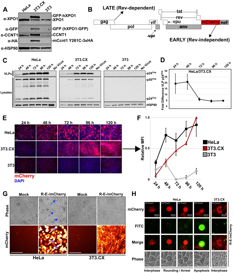
Cells derived from mice and other rodents exhibit profound blocks to HIV-1 virion production, reflecting species-specific incompatibilities between viral Tat and Rev proteins and essential host factors cyclin T1 (CCNT1) and exportin-1 (XPO1, also known as CRM1), respectively. To determine if mouse cell blocks other than CCNT1 and XPO1 affect HIV's postintegration stages, we studied HIV-1NL4-3 gene expression in mouse NIH 3T3 cells modified to constitutively express HIV-1-compatible versions of CCNT1 and XPO1 (3T3.CX cells). 3T3.CX cells supported both Rev-independent and Rev-dependent viral gene expression and produced relatively robust levels of virus particles, confirming that CCNT1 and XPO1 represent the predominant blocks to these stages. Unexpectedly, however, 3T3.CX cells were remarkably resistant to virus-induced cytopathic effects observed in human cell lines, which we mapped to the viral protein Vif and its apparent species-specific capacity to induce G2/M cell cycle arrest. Vif was able to mediate rapid degradation of human APOBEC3G and the PPP2R5D regulatory B56 subunit of the PP2A phosphatase holoenzyme in mouse cells, thus demonstrating that VifNL4-3's modulation of the cell cycle can be functionally uncoupled from some of its other defined roles in CUL5-dependent protein degradation. Vif was also unable to induce G2/M cell cycle arrest in other nonhuman cell types, including cells derived from nonhuman primates, leading us to propose that one or more human-specific cofactors underpin Vif's ability to modulate the cell cycle.IMPORTANCE Cells derived from mice and other rodents exhibit profound blocks to HIV-1 replication, thus hindering the development of a low-cost small-animal model for studying HIV/AIDS. Here, we engineered otherwise-nonpermissive mouse cells to express HIV-1-compatible versions of two species-specific host dependency factors, cyclin T1 (CCNT1) and exportin-1 (XPO1) (3T3.CX cells). We show that 3T3.CX cells rescue HIV-1 particle production but, unexpectedly, are completely resistant to virus-induced cytopathic effects. We mapped these effects to the viral accessory protein Vif, which induces a prolonged G2/M cell cycle arrest followed by apoptosis in human cells. Combined, our results indicate that one or more additional human-specific cofactors govern HIV-1's capacity to modulate the cell cycle, with potential relevance to viral pathogenesis in people and existing animal models.

Keywords: APOBEC3G; G2/M; HIV; PPP2R5D; Rev; Vif; cell cycle; cyclin T1; exportin-1; species specific.


Figures

FIG 1
FIG 1
Stable expression of mCcnt1-Y261C and hXPO1 is sufficient to rescue virus particle production in murine cells. (A) Western blot analysis comparing 3T3.CX cells that express mCcnt1 Y261C-3xHA and GFP-hXPO1 to permissive HeLa cells and the nonpermissive 3T3 parental control cell line. (B) Genomic layout of the NL4-3 strain HIV-1 reporter virus used in this study. In this virus, env and vpr genes are inactivated due to frameshift mutations, and the gene encoding red fluorescent protein mCherry was inserted into the nef locus, termed R-E-/mCherry. (C) HeLa, 3T3.CX, and 3T3 cells were infected with VSV-G-pseudotyped R-E-/mCherry virus at an MOI of ∼1 TU. Western blot analysis of supernatants and cell lysates harvested at 24-h time points as described in Materials and Methods. Gag species were detected using anti-p24Gag (CA) monoclonal antiserum and secondary antibodies conjugated to infrared fluorophores. Heat shock protein 90 (HSP90) was detected as a loading control. Peak rates of VLP production were observed between 24 and 48 hpi in HeLa cells (left), with peak rates of 3T3.CX cell production occurring later, at 48 to 72 hpi (middle). Little to no Gag/Gag-Pol expression was observed in parental 3T3 cells at any time point (right). (D) Fold differences in VLP p24Gag levels from panel C plotted over time, comparing infection of HeLa cells to that of 3T3.CX cells. Error bars represent the standard deviations from the means (n = 3); data show a representative example from three biological replicates. (E, F) mCherry expression was monitored using microscopy and quantified using a fluorescence spectrophotometer at 24-h time points. Bars, ∼100 μm. 3T3.CX cells exhibited robust mCherry expression after 72 hpi. Error bars represent the standard deviations from the means (n = 3); data show a representative example from three biological replicates. (G) Fluorescence microscopy of HeLa and 3T3.CX cells infected with R-E-/mCherry. Infected HeLa cells, but not 3T3.CX cells, exhibited extensive cell rounding at 72 hpi (arrows). Bars, ∼100 μm. (H) Still images from live-cell fluorescence microscopy of infected HeLa cells stained with a caspase 3/7 detection reagent (fluorescein isothiocyanate [FITC] channel). HeLa cells rounded at ∼30 hpi when infected with R-E-/mCherry and arrested for ∼10 to 20 h prior to undergoing apoptosis. In contrast, infected 3T3.CX cells were not observed to undergo cell rounding or apoptosis at any time point. The images in this panel can be seen in Movie S1.
FIG 2
FIG 2
HIV-1 Vif causes CPE and G2/M cell cycle arrest in HeLa cells but not in 3T3.CX cells. HeLa and 3T3.CX cells were infected with either Vif+ or ΔVif R-E-/CFP viruses. (A) Visualization of infected HeLa and 3T3.CX cells. Bars, ∼100 μm. For all CFP images, all CFP fluorescence was pseudocolored black on a white background to maximize contrast. Accordingly, uninfected cells or infected cells expressing CFP at or below background are shown as white. Only HeLa cells infected with a Vif+ virus underwent cell rounding (insets), while HeLa cells infected with ΔVif viruses maintained normal cell morphology despite infection. 3T3.CX cells maintained normal cell morphology regardless of which virus (Vif+ or ΔVif) was used to infect cells. (B) Both HeLa and 3T3.CX cells were infected with Vif+ R-E-/CFP or ΔVif R-E-/CFP as indicated, harvested, and analyzed using flow cytometry to quantify per-cell DNA content using propidium iodide staining. For flow histograms, the leftmost peak indicates DNA content prior to S phase (G1), with the rightmost peak demonstrating cells with duplicated DNA content (G2/M). The blue curve between the G1 and G2/M peaks represents the S phase. Quantification of G2/M status for each condition is presented on the right of the flow histograms. Infecting HeLa cells with Vif+ viruses induced G2/M cell cycle arrest, while ΔVif virus did not, consistent with the observed cell morphology changes presented in panel A. 3T3.CX cells were nonresponsive to either virus and did not undergo G2/M cell cycle arrest. Error bars represent the standard deviations from the means (n = 3). (C) Treating HeLa or 3T3.CX cells with 1 μM nocodazole for 24 h induced potent G2/M cell cycle arrest and cell rounding changes in both cell lines (rightmost images). Error bars represent the standard deviations from the means (n = 3).
FIG 3
FIG 3
Vif alone is sufficient to induce cell rounding and G2/M cell cycle arrest in HeLa cells but not in 3T3.CX cells. (A) Depiction of bicistronic retroviral vectors that express both codon-optimized Vif (CO-Vif) and cyan fluorescent protein (CFP) from reading frames separated by an internal ribosomal entry site (IRES). Three independent constructs were studied: CO-VifNL4-3 (induces G2/M arrest), C114S CO-VifNL4-3 (catalytically inactive, should not induce G2/M arrest), and CO-VifHXB2 (should not induce G2/M arrest). (B) Western blot analysis confirming that each construct expressed CO-Vif in addition to CFP, assayed in HeLa cells. (C) Visualization of HeLa cells and 3T3 cells transduced with each construct, separately. Bar, ∼100 μm. HeLa cells that expressed CO-VifNL4-3 underwent marked cell rounding (insets), while HeLa cells expressing C114S CO-VifNL4-3 and CO-VifHXB2 maintained normal cell morphology (compare mock [Vector] images to CO-Vif images). 3T3 cells, however, maintained normal cell morphology under every condition. Treatment with 1 μM nocodazole as a G2/M control induced cell rounding in both cell types. (D) Cell cycle analysis of HeLa and 3T3 cells using flow cytometry confirmed that 3T3 cells failed to undergo G2/M cell cycle arrest in the presence of CO-VifNL4-3 expression. Error bars represent the standard deviations from the means (n = 3).
FIG 4
FIG 4
Vif is capable of degrading hA3G, CBF-β, and PPP2R5D in both HeLa and 3T3 cells. (A) Model of the Vif-APOBEC3G E3 ubiquitin ligase complex based on that proposed by Letko et al. (95). In this model, Vif (cyan) is bound to elongin-C (orange), CBF-β (gray), and CUL5 (purple), with species-specific CUL5 residues near the Vif-binding interface highlighted in yellow. Elongin-B (red) is shown bound to elongin-C but does not bind to Vif directly. Amino acid sequences identifying mouse versus human orthologues of each factor are as indicated in the table. (B) HeLa and 3T3 cell lines stably expressing YFP-hA3G (yellow) were transduced to express increasing amounts of CO-VifNL4-3 as indicated. Images show YFP-hA3G degradation at 48 h postransduction, based on the disappearance of YFP fluorescence detected by microscopy using a 10× objective lens for ∼100 cells/field and measured using FIJI/ImageJ2 software. Bar, ∼100 μm. Insets show bright-field image of cells in order to illustrate the consistent cell density at 48 h. Note that YFP-hA3G is practically undetectable at the highest dose (800 μl) of Vif vector, consistent with efficient degradation. (C) Western blot analysis showing relative Vif, YFP-hA3G, CBFβ, and PPP2R5D levels for an experiment identical to that presented in panel B. HSP90 served as a negative control for Vif effects and also as a loading control. (D) Quantification of YFP-hA3G and PPP2R5D levels relative to HSP90 based on quantitative Western blotting (as shown in panel C) and confirming similar, dose-dependent degradation levels in either cell type. Error bars represent the standard deviations from the means (n = 3).
FIG 5
FIG 5
Vif-induced cell cycle arrest is species specific. (A) Images of cell lines derived from humans (HeLa, HOS, U2OS) or a nonhuman primate (COS7) and infected with either Vif+ or ΔVif R-E-/mCherry HIV-1 and visualized using a 10× objective lens at 3 days postinfection. Bar, ∼100 μm. Inset images highlight frequent observations of cell rounding and cytopathic effect. (B) Additional informative nonhuman cell lines derived from rodent (mouse L cells, Rat1, Chinese hamster ovary [CHO]), chiropteran (TB1), and avian (DF1) species and infected with Vif+ and ΔVif viruses as indicated. Images were collected as described for panel A and are shown for a later time point, 7 days postinfection, to demonstrate that infection by either virus was tolerated for all cell types relative to the HeLa cell control. Bar, ∼100 μm. Inset images highlight the few remaining HeLa cells in culture at 7 days postinfection with the Vif+ virus. (C) Quantification of mCherry mean fluorescence intensity (MFI) for whole-field imaging of the cells presented in panel A, demonstrating the loss of mCherry-expressing human cells at 6 days postinfection. Error bars represent the standard deviations from the means for three independent infections. For both panels C and D, the skull and crossbone symbols indicate high levels of cell death (as opposed to reductions to per-cell mCherry fluorescence). (D) Plot of mCherry MFI, as described for panel C, for whole-field imaging of the cells at 7 days postinfection presented in panel B. (E) Western blot analysis for cell lysates as described for Fig. 1E depicting Gag/Gag-Pol expression and cleavage profiles for HeLa and Cos7 cells infected with the indicated viruses and harvested at 6 days postinfection. Equivalent amounts of total protein were loaded into each well, thus confirming that Cos7 cells can tolerate and maintain infection with a Vif+ virus over multiple cell divisions, while infected HeLa cells are gradually lost from the culture. (F) Cell cycle analysis as described for Fig. 3, comparing HeLa and Cos7 cells transduced to express CO-VifNL4-3 in the absence of other viral factors. Notably, Cos7 cells were largely resistant to HIV-1 Vif-mediated G2/M cell cycle arrest. Error bars represent the standard deviations from the means (n = 3).
FIG 6
FIG 6
Proposed models for mouse cell resistance of HIV-1 Vif-mediated G2/M cell cycle arrest. We present two working models for Vif-mediated G2/M cell cycle arrest. (A) Vif is able to identify and ultimately degrade a yet-to-be identified substrate that regulates G2/M arrest in human cells. In mouse cells, this substrate either is lacking or does not interact with Vif. (B) In human cells, the recently identified PP2A subunit substrates are degraded by Vif to induce G2/M arrest, perhaps through modulation of aurora kinase activity, as proposed by Greenwood et al. (47). Vif can degrade PP2A subunits in mouse cells (Fig. 4), but this mechanism may be uncoupled from a downstream, human-specific regulatory factor.

Similar articles

Cited by

References

    1. Malim MH, Bieniasz PD. 2012. HIV restriction factors and mechanisms of evasion. Cold Spring Harb Perspect Med 2:a006940. doi:10.1101/cshperspect.a006940. - DOI - PMC - PubMed
    1. van Maanen M, Sutton RE. 2003. Rodent models for HIV-1 infection and disease. Curr HIV Res 1:121–130. doi:10.2174/1570162033352075. - DOI - PubMed
    1. Shida H. 2012. Role of nucleocytoplasmic RNA transport during the life cycle of retroviruses. Front Microbiol 3:179. doi:10.3389/fmicb.2012.00179. - DOI - PMC - PubMed
    1. Seay K, Qi X, Zheng JH, Zhang C, Chen K, Dutta M, Deneroff K, Ochsenbauer C, Kappes JC, Littman DR, Goldstein H. 2013. Mice transgenic for CD4-specific human CD4, CCR5 and cyclin T1 expression: a new model for investigating HIV-1 transmission and treatment efficacy. PLoS One 8:e63537. doi:10.1371/journal.pone.0063537. - DOI - PMC - PubMed
    1. Atchison RE, Gosling J, Monteclaro FS, Franci C, Digilio L, Charo IF, Goldsmith MA. 1996. Multiple extracellular elements of CCR5 and HIV-1 entry: dissociation from response to chemokines. Science 274:1924–1926. doi:10.1126/science.274.5294.1924. - DOI - PubMed

Publication types

MeSH terms