. 2018 Jan 10;8(1):230.
doi: 10.1038/s41598-017-18431-6.

Xeno-free pre-vascularized spheroids for therapeutic applications

Affiliations

Xeno-free pre-vascularized spheroids for therapeutic applications

E Bauman et al. Sci Rep. .

Abstract

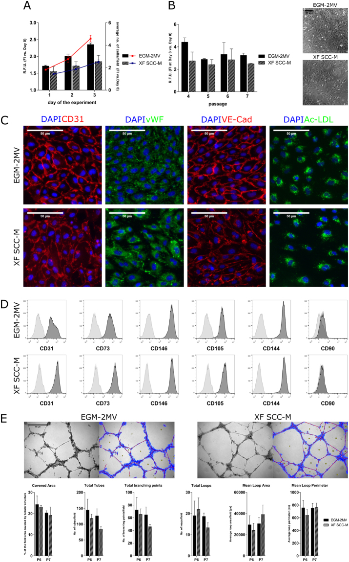
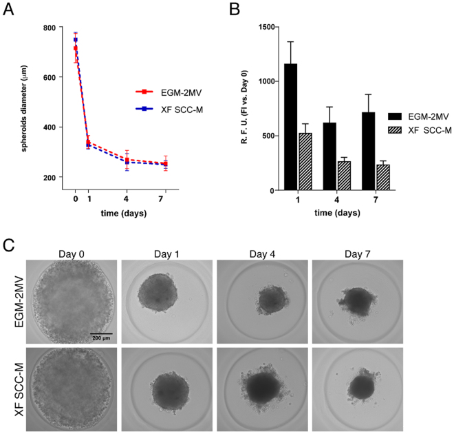
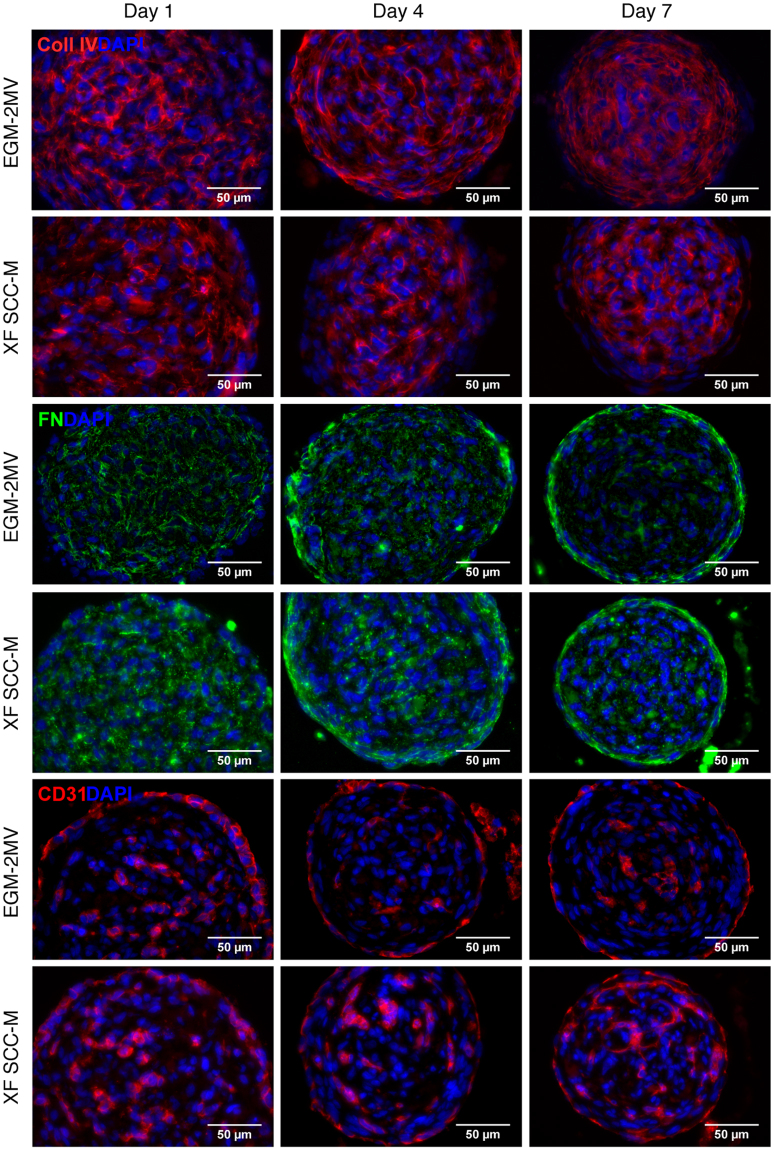
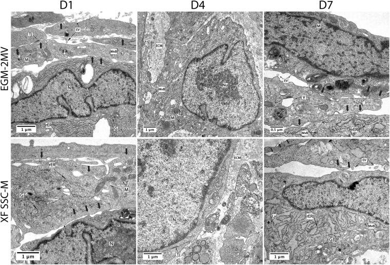
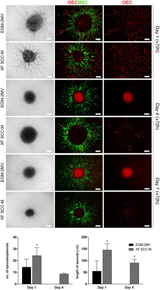
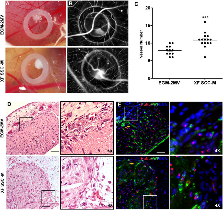
Spheroid culture has gained increasing popularity, arising as a promising tool for regenerative medicine applications. Importantly, spheroids may present advantages over single-cell suspensions in cell-based therapies (CT). Unfortunately, most growth media used for spheroid culture contain animal origin-components, such as fetal bovine serum (FBS). The presence of FBS compromises the safety of CT and presents economic and ethical constraints. SCC (supplement for cell culture) is a novel xeno-free (XF) industrial cell culture supplement, derived from well-controlled pooled human plasma and processed under good manufacturing practice rules. Here, we developed a XF SCC-based formulation for 2D-culture of outgrowth endothelial cells (OEC), and then used it for generating co-culture spheroids of OEC and mesenchymal stem cells (MSC). XF MSC-OEC spheroids were characterized in detail and compared to spheroids cultured in FBS-supplemented medium. XF spheroids presented comparable integrity, size and morphology as the reference culture. The use of both media resulted in spheroids with similar structure, abundant extracellular matrix deposition and specific patterns of OEC distribution and organization. Notably, XF spheroids presented significantly enhanced angiogenic potential, both in vitro (fibrin sprouting assay) and in vivo (CAM assay). These findings are particularly promising in the context of potential therapeutic applications.


Conflict of interest statement

The authors declare that they have no competing interests.

Figures

Figure 1
Figure 1
Optimization of the XF SCC-M in OEC culture. (A) OEC proliferation in the XF SCC-M. Cell growth of OEC in the control and XF conditions, expressed respectively in the metabolic activity levels and the rate of culture expansion measured by high throughput cell number quantification. (B) XF SCC-M formulation performance in terms of cell metabolic activity, analysed over the passages of OEC culture. Phase contrast images of OEC morphology in the control and XF formulation. (C) Representative images showing cellular localization of CD31, vWF, VE-Cad, and Ac-LDL in standardly and SCC-grown OEC. (D) Representative flow cytometry histograms of OEC cultured in the control medium and XF SCC-M showing reactivity with EC-characteristic marker molecules (right-shifted filled black curves compared with grey lined curves of the appropriate controls) and lack of reactivity with the negative CD90 marker. (E) Representative micrographs of the tubular-like structures formed in Matrigel, along with exemplary analyses of OEC grown in EGM-2MV and XF SCC-M. Quantification of characteristic parameters of the tubular structure derived from EGM-2MV and XF SCC-M-cultured cells.
Figure 2
Figure 2
Generation of MSC-OEC spheroids under XF conditions. (A) Brigthfield images of MSC-OEC spheroids compaction along time. (B) Metabolic activity of the spheroids in culture. (C) Morphology of MSC-OEC spheroids over 7-day culture (scale bar 100 μm).
Figure 3
Figure 3
Structure/internal organization of the MSC-OEC spheroids under XF conditions. Representative images showing deposition of ECM proteins (collagen type IV and fibronectin) and cellular organization of the spheroids along the culture, respectively in the control and SCC-based medium immunostaining of collagen type IV (red), FN (green), CD31 (red) counterstained with DAPI (blue). Images were taken with a 40X objective. Scale bar 50 μm.
Figure 4
Figure 4
Transmission electron microscopy (TEM) analysis of MSC-OEC spheroids external layers ultrastructure. Images depict elongated cells (arrows); accumulation of ECM – extracellular matrix components Tj – tight junctions and different organelles including N – nuclei; Nu – nucleolus; M – mitochondria; G – golgi apparatus; ER – endoplasmic reticulum; rER – rough endoplasmic reticulum; Ly- lysosomes; V- vacuoles; MvB- multivesicular bodies and EV- extracellular vesicles. Images were obtained with 12 K and 15 K and 35k magnification.
Figure 5
Figure 5
Fibrin-based 3D angiogenesis assay. Representative Brightfield and fluorescence images of sprouting MSC-OEC spheroids, cultured in normal and XF conditions for periods of respectively 1, 4, and 7 days; brought under the assay conditions for 72 h. OEC were marked with DiI-Ac-LDL and MSC were labelled with a CellTracker Green Dye. Scale bars correspond to 100 μm. Graphs represent quantitative analyses of the average numbers of sprouts per spheroid and their length, respectively under normal and XF conditions (n = 7, Mann-Whitney test was used to compare the two groups).
Figure 6
Figure 6
In vivo CAM assay. The angiogenic potential of spheroids in both conditions was tested using chick embryonic membrane. (A) Brightfield images of spheroids inside the CAM O-ring; (B) photomicrographs acquired for counting newly formed vessels; (C) Angiogenic effect of spheroids grown in the standard versus XF conditions, expressed by number of newly formed vessels (n ≥ 12, two independent experiments, ***p < 0.0004, Mann-Whitney test was used for statistical comparison). (D) HE staining of CAM interface and spheroids after 72 h incubation time (Scale bar 100 μm). (E) Immunofluorescence of equivalent sections represented in D (Scale bar 50 μm). vWF (green) was used to access endothelial organization, HuNu (red) to discriminate human nuclei and DAPI (blue) for total nuclei. In images (D) and (E), insets (and respective 4× magnifications, depicted on the right) highlight the direct contact between spheroids and the CAM. Yellow arrows are pointing to organized OEC inside the compacted spheroids.

References

    1. Bianco P, et al. The meaning, the sense and the significance: translating the science of mesenchymal stem cells into medicine. Nat med. 2013;19:35–42. doi: 10.1038/nm.3028. - DOI - PMC - PubMed
    1. Belema-Bedada F, Uchida S, Martire A, Kostin S, Braun T. Efficient homing of multipotent adult mesenchymal stem cells depends on FROUNT-mediated clustering of CCR2. Cell stem cell. 2008;2:566–575. doi: 10.1016/j.stem.2008.03.003. - DOI - PubMed
    1. Mao F, et al. Mesenchymal stem cells and their therapeutic applications in inflammatory bowel disease. Oncotarget. 2017;23:38008–38021. - PMC - PubMed
    1. Laschke MW, Menger MD. Life is 3D: boosting spheroid function for tissue engineering. Trends Biotechnol. 2017;35:133–144. doi: 10.1016/j.tibtech.2016.08.004. - DOI - PubMed
    1. Rouwkema J, Khademhosseini A. Vascularization and angiogenesis in tissue engineering: beyond creating static networks. Trends Biotechnol. 2016;34:733–745. doi: 10.1016/j.tibtech.2016.03.002. - DOI - PubMed

Publication types

MeSH terms