. 2018 Jan 10;8(1):387.
doi: 10.1038/s41598-017-18672-5.

Respiratory Syncytial Virus Infection Changes Cargo Composition of Exosome Released from Airway Epithelial Cells

Affiliations

Respiratory Syncytial Virus Infection Changes Cargo Composition of Exosome Released from Airway Epithelial Cells

Harendra Singh Chahar et al. Sci Rep. .

Abstract

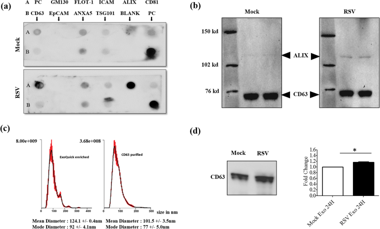
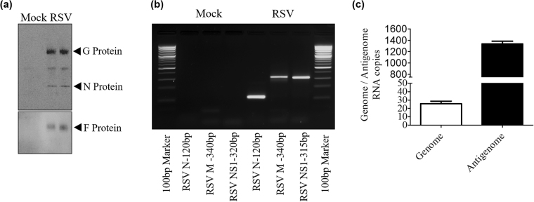
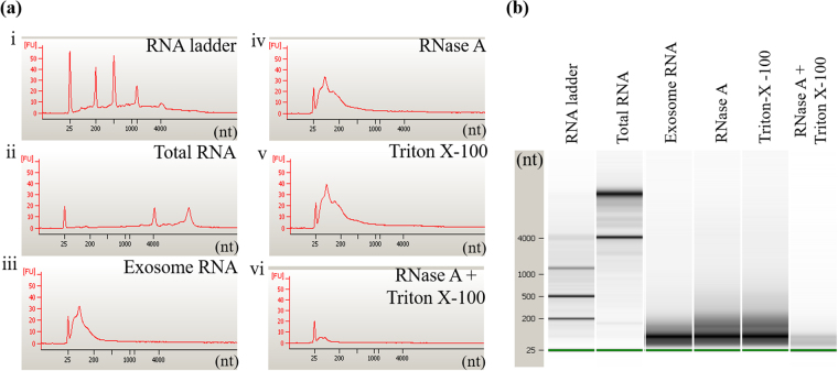
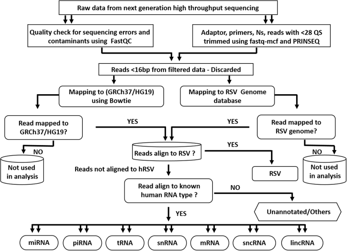
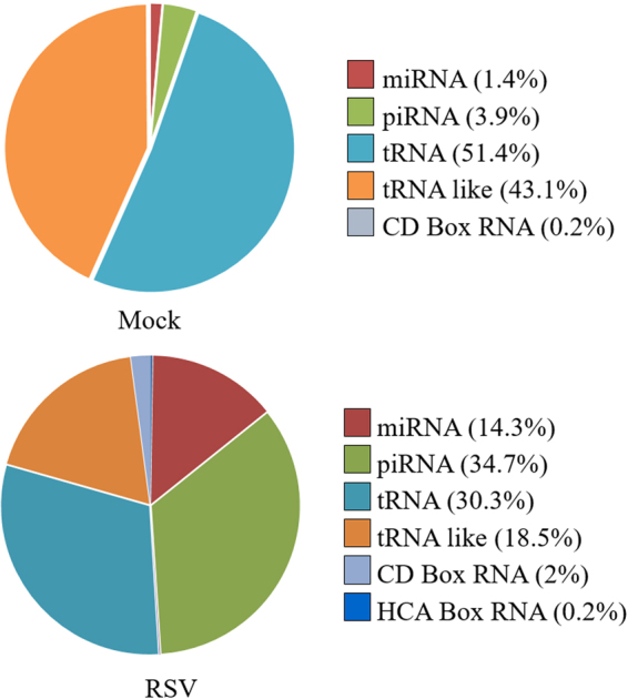
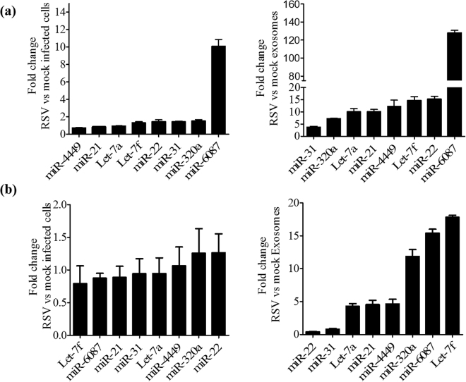
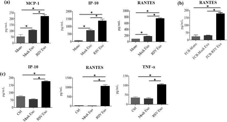
Exosomes are microvesicles known to carry biologically active molecules, including RNA, DNA and proteins. Viral infections can induce profound changes in exosome composition, and exosomes have been implicated in viral transmission and pathogenesis. No information is current available regarding exosome composition and function during infection with Respiratory Syncytial Virus (RSV), the most important cause of lower respiratory tract infections in children. In this study, we characterized exosomes released from RSV-infected lung carcinoma-derived A549 cells. RNA deep sequencing revealed that RSV exosomes contain a diverse range of RNA species like messenger and ribosomal RNA fragments, as well as small noncoding RNAs, in a proportion different from exosomes isolated from mock-infected cells. We observed that both RNA and protein signatures of RSV were present in exosomes, however, they were not able to establish productive infection in uninfected cells. Exosomes isolated from RSV-infected cells were able to activate innate immune response by inducing cytokine and chemokine release from human monocytes and airway epithelial cells. These data suggest that exosomes may play an important role in pathogenesis or protection against disease, therefore understating their role in RSV infection may open new avenues for target identification and development of novel therapeutics.


Conflict of interest statement

The authors declare that they have no competing interests.

Figures

Figure 1
Figure 1
Diagram of exosome isolation and characterization. Exosomes were isolated using ExoQuick reagent from conditioned cell culture supernatants and subjected to CD63 immuno-magnetic selection for recovery of highly pure exosomes. Immuno-purified exosomes were then characterized by western blot, using an exosome marker antibody array, and by nanoparticle tracking analysis.
Figure 2
Figure 2
Characterization of purified exosomes. (a) Characterization of equal amounts of purified exosomes by protein marker antibody array. The exosome marker array detects 8 known exosome markers namely – CD63, Immunoglobulin superfamily, member 8 (CD81), Programmed cell death 6 interacting protein (ALIX), Flotillin 1 (FLOT1), Intercellular adhesion molecule 1 (ICAM1), Epithelial cell adhesion molecule (EpCam), Annexin A5 (ANXA5) and Tumor susceptibility gene 101 (TSG101). Cis-Golgi matrix protein marker GM130 serves as control to monitor cellular contamination in exosome preparations. PC stands for positive control. (b) Western blot of equal amounts of CD63-purified exosomes using the exosome markers ALIX and CD63. (c) Absolute size determination and quantification of RSV exosomes either reagent enriched or CD63-purified by NanoSight LM10 analysis. The particles were tracked and sized based on Brownian motion and the diffusion coefficient. The mean and mode diameter of exosomes particles are shown. The absolute count of exosome particles was determined and expressed as particles/ml. (d) Exosomes were enriched from 24 h cell supernatants and equal volumes were analyzed for CD63 expression by Western blot assay. Right panel represents densitometric analysis of three independent experiments. ‘*’ Indicates a statistically significant difference (P value < 0.05) comparing RSV exosomes versus mock.
Figure 3
Figure 3
RSV viral RNA and protein content in exosomes. (a) Western blot of protein lysates of exosomes using a polyclonal antibody against RSV. (b) RSV N, M and NS1 gene amplification by RT-PCR from CD63-purified exosome RNA. (c) qRT-PCR amplification of genomic and antigenomic RNA present in RSV exosomes.
Figure 4
Figure 4
Exosome RNA cargo length profile. (a) Electropherograms and (b) gel images of RNA extracted from exosomes untreated or treated with RNase A in the presence or absence of Triton X-100 and run on the Agilent Bioanalyzer to determine size of RNA fragments.
Figure 5
Figure 5
Next generation high-throughput RNA sequencing analysis pipeline flowchart. The flowchart demonstrates how the next generation sequencing data was analyzed. In brief, raw sequencing reads were quality checked for sequencing errors and contaminants using FastQC. Adapter sequences, primers, Ns, and reads with quality score below 28 were trimmed using fastq-mcf of ea-utils and PRINSEQ. Reads < 16 bp after trimming were discarded. Pseudo single-end reads were mapped to the human genome using bowtie. Raw read counts were calculated for known gene categories including ncRNAs, antisense transcripts, coding and intronic regions of mRNAs, and repeats. Annotations of known genes were retrieved from miRBase release 20, NCBI RefSeq, Human lincRNA Catalog, and UCSC Genome Browser. hRSV- Human Respiratory Syncytial Virus, miRNA- microRNA, piRNA- piwi interacting RNA, tRNA- transfer RNA, snRNA- small nucleolar RNA, mRNA-messenger RNA, sncRNA-small noncoding RNA, lincRNA- long intergenic noncoding RNA.
Figure 6
Figure 6
Graphic representation of the relative abundance of small ncRNAs from exosomes.
Figure 7
Figure 7
Validation of miRNA expression in cells and exosomes. RNA extracted from A549 cells (a) or SAE cells (b) mock- and RSV-infected for 24 h (left panels) and from mock and RSV exosomes (right panels) was subjected to miRNA analysis by RT-PCR. Fold changes in miRNA expression were determined by the 2-ΔΔCt method and represent mean ± SD normalized to small-nucleolar RNA U6 (RNU6).
Figure 8
Figure 8
Proinflammatory mediator secretion by monocytes and A549 cells in response to exosome stimulation. Human monocytes were stimulated with mock or RSV exosomes and harvested 24 h later to collect cell supernatants to measure cytokine and chemokine secretion by cytokine multiplex assay (a) or ELISA (b). Similar experiment was performed in A549 cells (c). Data are expressed as mean ± SEM, and ‘*’ indicates a statistically significant difference (P value < 0.05).

References

    1. Harding C, Heuser J, Stahl P. Receptor-mediated endocytosis of transferrin and recycling of the transferrin receptor in rat reticulocytes. J. Cell Biol. 1983;97:329–339. doi: 10.1083/jcb.97.2.329. - DOI - PMC - PubMed
    1. Harding C, Heuser J, Stahl P. Endocytosis and intracellular processing of transferrin and colloidal gold-transferrin in rat reticulocytes: demonstration of a pathway for receptor shedding. Eur. J. Cell Biol. 1984;35:256–263. - PubMed
    1. Johnstone RM, Adam M, Hammond JR, Orr L, Turbide C. Vesicle formation during reticulocyte maturation. Association of plasma membrane activities with released vesicles (exosomes) J. Biol. Chem. 1987;262:9412–9420. - PubMed
    1. Zech D, Rana S, Buchler MW, Zoller M. Tumor-exosomes and leukocyte activation: an ambivalent crosstalk. Cell Commun. Signal. 2012;10:37. doi: 10.1186/1478-811X-10-37. - DOI - PMC - PubMed
    1. Thery C, et al. Molecular characterization of dendritic cell-derived exosomes. Selective accumulation of the heat shock protein hsc73. J. Cell Biol. 1999;147:599–610. doi: 10.1083/jcb.147.3.599. - DOI - PMC - PubMed

Publication types

MeSH terms