. 2018 Jan 10;8(1):237.
doi: 10.1038/s41598-017-18652-9.

Blimp-1/PRDM1 is a critical regulator of Type III Interferon responses in mammary epithelial cells

Affiliations

Blimp-1/PRDM1 is a critical regulator of Type III Interferon responses in mammary epithelial cells

Salah Elias et al. Sci Rep. .

Abstract

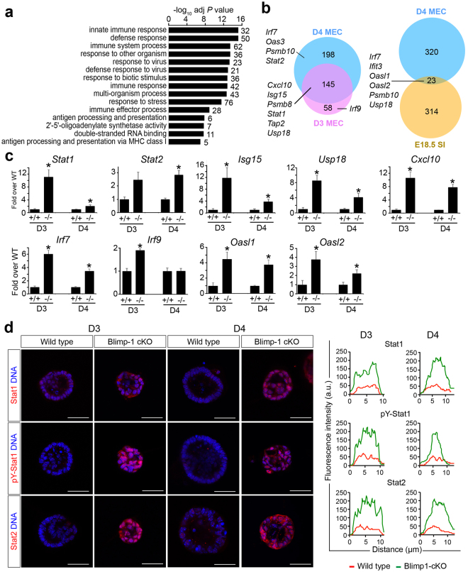
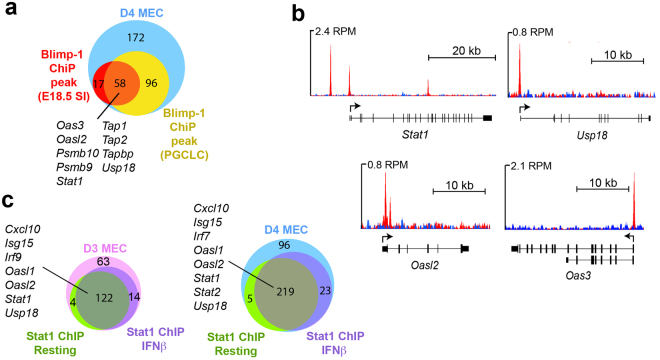
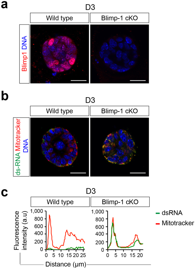
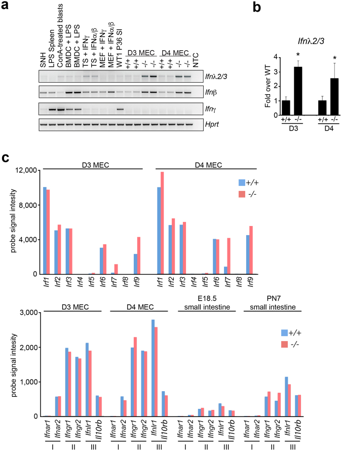
The transcriptional repressor Blimp-1 originally cloned as a silencer of type I interferon (IFN)-β gene expression controls cell fate decisions in multiple tissue contexts. Conditional inactivation in the mammary gland was recently shown to disrupt epithelial cell architecture. Here we report that Blimp-1 regulates expression of viral defense, IFN signaling and MHC class I pathways, and directly targets the transcriptional activator Stat1. Blimp-1 functional loss in 3D cultures of mammary epithelial cells (MECs) results in accumulation of dsRNA and expression of type III IFN-λ. Cultures treated with IFN lambda similarly display defective lumen formation. These results demonstrate that type III IFN-λ profoundly influences the behavior of MECs and identify Blimp-1 as a critical regulator of IFN signaling cascades.


Conflict of interest statement

The authors declare that they have no competing interests.

Figures

Figure 1
Figure 1
Transcriptional profiling experiments reveal up-regulated expression of anti-viral defense genes in Blimp-1 cKO mammary epithelial cell (MEC) cultures. (a) Significantly enriched GO terms among up-regulated genes in Day 4 Blimp-1 cKO MECs as determined by WebGestault. (b) Comparisons of up-regulated transcripts in Blimp-1 cKO MEC 3D cultures at Day 3 and Day 4 with those up-regulated in Blimp-1 cKO E18.5 small intestine identify components of viral defense, IFN signaling cascades and MHC class I antigen presentation pathways. (c) qPCR validation of transcriptional changes in cKO (−/−) relative to wild type (+/+). *P value < 0.05 (unpaired Student’s t-test). Data represents mean ± SEM of 5 samples per group. (d) Dramatically increased Stat1, pY-Stat1 and Stat2 expression in Blimp-1cKO 3D cultures. Representative line scan-analysis (representative fluorescence intensity, minimum 20 cells/genotype analyzed) confirms increased levels and nuclear localization of Stat1, pY-Stat1, and Stat2 in Blimp-1 cKO MECs. Scale bars: 50 μm.
Figure 2
Figure 2
Comparisons with Blimp-1 and Stat1 ChIP-Seq datasets identifies direct target genes. (a) The subset of up-regulated genes with proximal Blimp-1 ChIP peaks includes Stat1, Oas family members, Usp18, and IFN-inducible components of the MHC class I antigen presentation pathway. (b) Blimp-1 binding at Stat1, Usp18, Oasl2, and Oas3 genes (GSE66069). Positions of the TSS and direction of transcription are indicated by the arrows. (c) The majority of up-regulated genes contain proximal Stat1 ChIP-Seq peaks.
Figure 3
Figure 3
Blimp-1 inactivation in mammary epithelial cells results in accumulation of dsRNA. (a) Treatment with 4OHT activates Cre recombinase and eliminates Blimp-1 expression in mutant MEC 3D cultures. (b) Representative images of wild type and Blimp-1 cKO MEC 3D cultures co-stained for dsRNA using the J2 mAb and mitotracker to localize mitochondria. (c) Representative line scan-analysis (relative fluorescence intensity, minimum 20 cells/genotype analyzed) confirms increased levels of dsRNA and mitochondrial co-localization. Scale bars: 50 μm.
Figure 4
Figure 4
Blimp-1 functional loss activates type III IFN-λ expression in mammary epithelial cells. (a) RT-PCR analysis demonstrates induction of Type III λ and Type I β but not Type II γ IFN transcripts in Blimp-1 cKO MEC 3D cultures. Abbreviations: +/+, Wild type; −/−, Blimp-1 cKO; BMDC, bone marrow-derived dendritic cells; LPS, Lipopolysaccharide; ConA, Concanavalin; A SNH, STO cell line; MEF, mouse embryonic fibroblasts; TS, trophoblast stem cell; P36 SI, postnatal day 36 small intestine. Cropped PCR gel images shown. For uncropped gel images see Supplementary Figure S2. (b) qPCR validation of increased type III IFN-λ2/3 transcription. *P value < 0.05 (unpaired Student’s t-test). Data represents mean ± SEM of 4 samples per group. (c) Mammary epithelial cell 3D cultures robustly express multiple Irf family members and the type III IFN lambda receptor and as determined by average microarray probe signal intensity. Blimp-1 cKO MEC 3D cultures express increased levels of Irf7 (see also Supplementary Table S1). For comparison, microarray probe signals from embryonic (E) day 18.5 and postnatal (P) day 7 wild type and Blimp-1 cKO small intestine datasets corresponding to NCBI GEO accession numbers GSE29658 and GSE30556 respectively, are shown.
Figure 5
Figure 5
Disturbances caused by IFNλ treatment closely resemble those observed in Blimp-1 cKO 3D MEC cultures. (a) Phase contrast images of D6 MEC 3D cultures treated with recombinant type III IFN-λ2 reveal defective lumen formation and maturation. No noticeable changes were observed in the presence of Type II IFN-γ, whereas treatment with Type I IFN-β resulted in an intermediate phenotype. Bar graphs compare the percentages of acini with normal lumen and average acini diameters observed for Blimp-1 cKO vs wild type MEC cultures treated with recombinant IFNs. **P value < 0.01, ***P value < 0.001, relative to control (cKO vs WT - unpaired Student’s t-test, inter-treatment comparisons - ANOVA followed by Tukey post-hoc test). Data represents mean ± SEM of 282 (WT), 305 (cKO), 134 (Control), 112 (IFN-λ2), 145 (IFN-β) and 127 (IFN-γ) acini (normal lumen formation) and 104 (WT), 114 (cKO), 134 (Control), 112 (IFN-λ2), 145 (IFN-β) and 127 (IFN-γ) acini (diameter analysis). (b) Immunostaining demonstrates dramatically increased Stat1, pY-Stat1, and Stat2 expression induced by treatment with type III IFN-λ. Representative line scan-analysis (relative fluorescence intensity, in arbitrary units, a.u., minimum 20 cells per group analyzed). Scale bars: 50 μm.

References

    1. Keller AD, Maniatis T. Identification and characterization of a novel repressor of beta-interferon gene expression. Genes Dev. 1991;5:868–879. doi: 10.1101/gad.5.5.868. - DOI - PubMed
    1. Turner CA, Jr., Mack DH, Davis MM. Blimp-1, a novel zinc finger-containing protein that can drive the maturation of B lymphocytes into immunoglobulin-secreting cells. Cell. 1994;77:297–306. doi: 10.1016/0092-8674(94)90321-2. - DOI - PubMed
    1. Bikoff EK, Morgan MA, Robertson EJ. An expanding job description for Blimp-1/PRDM1. Curr Opin Genet Dev. 2009;19:379–385. doi: 10.1016/j.gde.2009.05.005. - DOI - PubMed
    1. Vincent SD, et al. The zinc finger transcriptional repressor Blimp1/Prdm1 is dispensable for early axis formation but is required for specification of primordial germ cells in the mouse. Development. 2005;132:1315–1325. doi: 10.1242/dev.01711. - DOI - PubMed
    1. Ohinata Y, et al. Blimp1 is a critical determinant of the germ cell lineage in mice. Nature. 2005;436:207–213. doi: 10.1038/nature03813. - DOI - PubMed

Publication types

Substances