

Cerebellar grey matter modifications in lower limb amputees not using prosthesis
- PMID: 29321625
- PMCID: PMC5762812
- DOI: 10.1038/s41598-017-18772-2
Cerebellar grey matter modifications in lower limb amputees not using prosthesis
Abstract
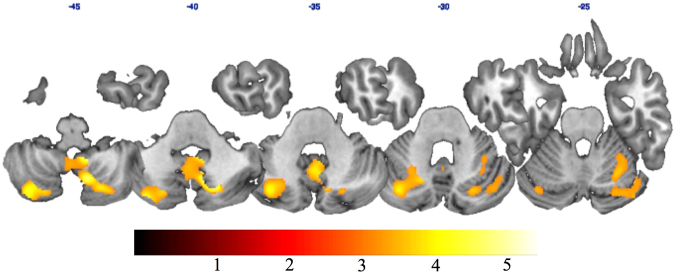
Plastic brain changes following peripheral deafferentation, in particular those following limb amputations, are well-documented, with significant reduction of grey matter (GM) in the sensory-motor cerebral areas representing the amputated limb. However, few studies have investigated the role played by the use of a prosthesis in these structural brain modifications. Here we hypothesized that using a functional prosthesis that allows individuals to perform actions may reduce grey matter reduction. We investigated the brain structural reorganization following lower limb amputation by using a Voxel Based Morphometry (VBM) analysis of structural magnetic resonance imaging (MRI) in 8 right-handed individuals with lower limb amputation (LLA) fitted with prostheses (LLAwp), compared to 6 LLA who had never used a prosthesis (LLAnp). 14 age-matched healthy controls were also enrolled (HC). We did not find any significant effect when comparing LLAwp and HC. However we found a decreased GM volume in the bilateral cerebellum in LLAnp compared with HC. These results suggest that prosthesis use prevents GM decrease in the cerebellum after lower limb amputation.
Conflict of interest statement
The authors declare that they have no competing interests.
Figures

References
Publication types
MeSH terms
LinkOut - more resources
Full Text Sources
Other Literature Sources
Medical
Molecular Biology Databases
