

Review
doi: 10.1038/s41388-017-0045-7.
Epub 2018 Jan 11.
Epidermal growth factor receptor and EGFRvIII in glioblastoma: signaling pathways and targeted therapies
Affiliations
- PMID: 29321659
- PMCID: PMC5860944
- DOI: 10.1038/s41388-017-0045-7
Item in Clipboard
Review
Epidermal growth factor receptor and EGFRvIII in glioblastoma: signaling pathways and targeted therapies
Oncogene.
2018 Mar.
Abstract
Amplification of epidermal growth factor receptor (EGFR) and its active mutant EGFRvIII occurs frequently in glioblastoma (GBM). While EGFR and EGFRvIII play critical roles in pathogenesis, targeted therapy with EGFR-tyrosine kinase inhibitors (TKIs) or antibodies has only shown limited efficacy in patients. Here we discuss signaling pathways mediated by EGFR/EGFRvIII, current therapeutics, and novel strategies to target EGFR/EGFRvIII-amplified GBM.
Conflict of interest statement
The authors declare no conflict of interest.
Figures
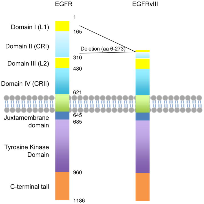
EGFR is a transmembrane tyrosine kinase receptor. The extracellular region includes four domains, L1, CR1, L2 and CR2. L1 and L2 are Leucine-rich domains that directly bind ligands. EGFRvIII is with the deletion of almost the entire L1 and CR1 domains, resulting in deficiency in ligand binding. The transmembrane and intracellular regions of EGFR and EGFRvIII are identical.
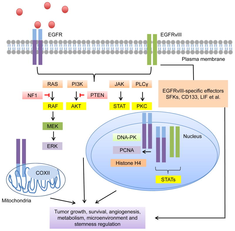
EGFR and EGFRvIII are able to transduce signals via classic RTK pathways including the RAS/RAF/MEK/ERK pathway, the PI3K/AKT pathway, the JAK/STAT pathway and the PKC pathway. The function of mitochondrial EGFR was also reported. Mitochondrial EGFR effector includes COXII. EGFR and EGFRvIII can also localize to the nucleus to activate a group of transcription factors and proteins involved in DNA damage responses, such as DNA-PK, PCNA, histone H4 and STATs. EGFRvIII has some unique signaling effectors. Activation of these signaling pathways and effector molecules together promote the fitness of the tumors.
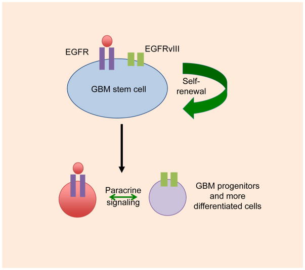
Rare cells in human GBM tumors that co-amplify EGFR and EGFRvIII show co-expression, with co-expression of EGFR and EGFRvIII driving malignancy more robustly, as compared to cells expressing EGFR or EGFRvIII alone. Most cells in human GBM tumors that co-amplify EGFR and EGFRvIII express predominantly EGFR or EGFRvIII, and cells expressing EGFRvIII can signal in a paracrine manner to cells expressing EGFR. A possible model incorporating these features is shown. See text for details.
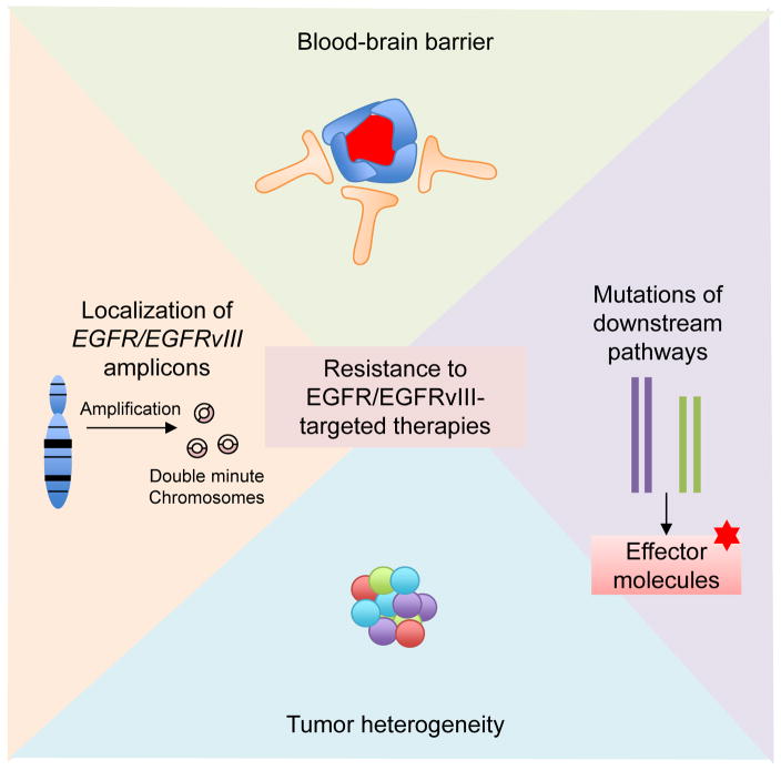
Factors contributing to resistance to EGFR/EGFRvIII inhibition include blood-brain barrier penetrance (i.e. many antibodies and chemicals cannot across the blood-brain barrier), mutations of signaling molecules downstream of EGFR/EGFRvIII (i.e. PTEN mutation and NF1 mutation, which maintain activation of downstream pathways despite upstream inhibition), tumor heterogeneity (distinct tumor cells can harbor different mutations or receptor kinase amplification, interaction between tumor cells and stromal cells in the tumor microenvironment) and extrachromosomal localization of EGFR and EGFRvIII amplicons (facilitating the cells’ ability to evade EGFR inhibitors).
References
-
- Cloughesy TF, Cavenee WK, Mischel PS. Glioblastoma: from molecular pathology to targeted treatment. Annual review of pathology. 2014;9:1–25. - PubMed
-
- Louis DN, Perry A, Reifenberger G, von Deimling A, Figarella-Branger D, Cavenee WK, et al. The 2016 World Health Organization Classification of Tumors of the Central Nervous System: a summary. Acta neuropathologica. 2016;131:803–820. - PubMed
-
- Ohgaki H, Kleihues P. The definition of primary and secondary glioblastoma. Clinical cancer research : an official journal of the American Association for Cancer Research. 2013;19:764–772. - PubMed
Publication types
MeSH terms
Substances
Grants and funding
LinkOut - more resources
Full Text Sources
Other Literature Sources
Medical
Molecular Biology Databases
Research Materials
Miscellaneous
