. 2017 Dec 13:11:119.
doi: 10.3389/fnana.2017.00119. eCollection 2017.

The Superior Fronto-Occipital Fasciculus in the Human Brain Revealed by Diffusion Spectrum Imaging Tractography: An Anatomical Reality or a Methodological Artifact?

Affiliations

The Superior Fronto-Occipital Fasciculus in the Human Brain Revealed by Diffusion Spectrum Imaging Tractography: An Anatomical Reality or a Methodological Artifact?

Yue Bao et al. Front Neuroanat. .

Abstract

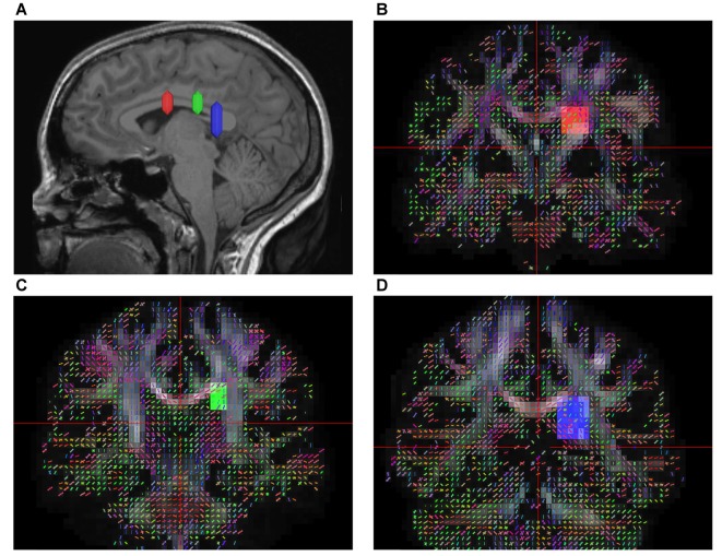
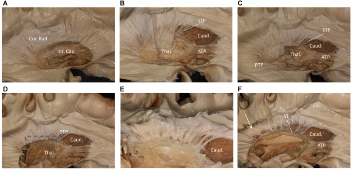
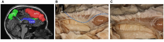
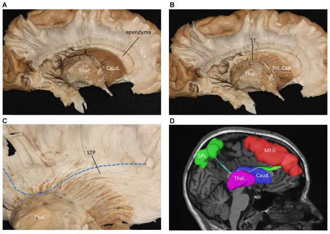
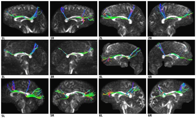
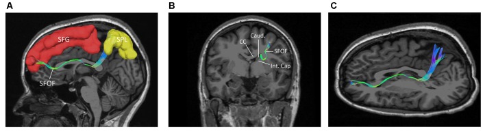
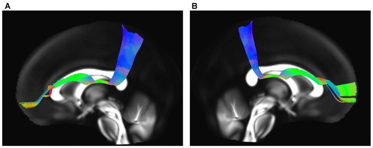
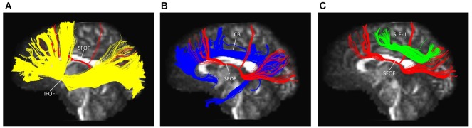
The existence of the superior fronto-occipital fasciculus (SFOF) in the human brain remains controversial. The aim of the present study was to clarify the existence, course, and terminations of the SFOF. High angular diffusion spectrum imaging (DSI) analysis was performed on six healthy adults and on a template of 842 subjects from the Human Connectome Project. To verify tractography results, we performed fiber microdissections of four post-mortem human brains. Based on DSI tractography, we reconstructed the SFOF in the subjects and the template from the Human Connectome Project that originated from the rostral and medial parts of the superior and middle frontal gyri. By tractography, we found that the fibers formed a compact fascicle at the level of the anterior horn of the lateral ventricle coursing above the head of caudate nucleus, medial to the corona radiate and under the corpus callosum (CC), and terminated at the parietal region via the lower part of the caudate nucleus. We consider that this fiber bundle observed by tractography is the SFOF, although it terminates mainly at the parietal region, rather than occipital lobe. By contrast, we were unable to identify a fiber bundle corresponding to the SFOF in our fiber dissection study. Although we did not provide definite evidence of the SFOF in the human brain, these findings may be useful for future studies in this field.

Keywords: diffusion spectrum imaging; fiber dissection; superior fronto-occipital fasciculus; tractography; white matter.


Figures

Figure 1
Figure 1
In vivo fiber tractography of the superior fronto-occipital fasciculus (SFOF). (A) Sagittal plane; lateral view of the hemisphere showing the rostrocaudal level of the three region of interests (ROIs), the three ROIs were located at the level of the anterior commissure, the thalamus and the pineal. (B–D) Coronal view on the QA map; all three ROIs were located under the corpus callosum (CC), lateral to the lateral ventricle and medial to the internal capusule.
Figure 2
Figure 2
Step by step fiber dissection from lateral to medial of right hemisphere. (A) The arcuate fasciculus (AF), superior longitudinal fasciculus (SLF) II and putamen were removed, the internal capsule was exposed. (B) After removing the anterior part of internal capsule, the head of caudate nucleus, thalamus, anterior thalamus peduncle and superior thalamus peduncle were exposed. (C) The anterior, superior and posterior thalamic peduncle. (D) Further removal of the internal capsule and thalamus, we could exposed the most medial part of thalamus peduncle. (E) Close-up and bottom to up view of the dotted rectangle in (D), it is clear that lots of fiber bundles reach thalamus. (F) After removal of the whole thalamus, some fibers as the white arrow shows coming from the parietal region have a close relationship to stria terminalis (ST). Int. Cap, internal capsule; Cor. Rad, corona radiate; Thal, thalamus; Caud, caudate nucleus; STP, superior thalamus peduncle; ATP, anterior thalamus peduncle; PTP, posterior thalamus peduncle; ST, stria terminalis.
Figure 3
Figure 3
(A) Fiber tractography of the SFOF (subject 3, right hemisphere); lateral view of the left SFOF, we found fibers originated from MFG curved around the upper part of the head of caudate nucleus and coursed to SPL. (B) The fibers related to ST. (C) From (C) to (B), we did not find some fiber bundles coursed above the head of caudate nucleus and connected the parietal region as the fiber tractography (dotted line) showed. But we did find some fibers interconnected the parietal lobe with the bottom part of caudate head via stria ternimalis as the white arrow showed. MFG, middle frontal gyrus; SPL, superior parietal lobe; caud, caudate; ST, stria terminalis.
Figure 4
Figure 4
Fiber dissection from medial to lateral of left hemisphere. (A) Caudate nucleus and thalamus were exposed. (B) After removal of the caudate nucleus, internal capsule and ST were exposed. (C) After removal of the ependyma, anterior portion of the subcallosal stratum and the radiation of the CC, we found the superior thalamus peduncle which came from the frontal lobe and reach the superior part of the thalamus. (D) Fiber tractography of the SFOF (subject 3, right hemisphere). We did not find an affirmative bundle of fibers interconnect the frontal lobe with parietal region as the dotted line in (C) and the SFOF in (D) showed. Thal, thalamus; Caud, caudate nucleus; Int. Cap, internal capsule; ST, stria terminalis; STP, superior thalamus peduncle; MFG, middle frontal gyrus; SPL, superior parietal lobe.
Figure 5
Figure 5
(A) Above the head of the caudate and medial to the internal capsule we can see some fibers come from frontal lobe. (B) Further dissection shows the fibers come from frontal region and bend inferiorly to the thalamus make up the STP. Cor. Rad, coronal radiation; Int. Cap, internal capsule; Thal, thalamus; STP, superior thalamus peduncle.
Figure 6
Figure 6
Diffusion spectrum imaging (DSI) tractography of the SFOF in 12 hemispheres of six subjects on sagittal view. Order: subject 1, 2, 3, 4, 5, 6. Left and right SFOF had a similar location, shape and trajectory in all 12 hemispheres. L, left; R, right.
Figure 7
Figure 7
The trajectory and cortical endpoints of the SFOF (subject-specific approach with its T1-weighted MPRAGE image; subject 3, left hemisphere). (A) Sagittal plane; lateral view of the left SFOF. SFOF originated from the superior frontal gyrus and terminated at the superior parietal lobe. (B) In the coronal section, the left SFOF located superolateral to the head of caudate nucleus, under the CC and medial to the internal capsule. (C) Combined axial and sagittal view, the left SFOF originated from the SFG and ran posteriorly lateral to the lateral ventricle, and then it ran postero-superiorly and finally terminated in the superior parietal lobule. SFG, superior frontal gyrus; MFG, middle frontal gyrus; SPL, superior parietal lobe; CC, corpus callosum; Caud, caudate nucleus; Int. Cap, internal capsule.
Figure 8
Figure 8
In vivo fiber tractography of the SFOF on Human Connectome Project (HCP-842) template. (A,B) SFOF in HCP-842 template and the HCP-842 template showed similar results to the subject-specific findings. L, left; R, right.
Figure 9
Figure 9
Spatial relationship of the SFOF with the adjacent association tracts. (A) Sagittal view showed the SFOF located above the inferior fronto-occipital fasciculus (IFOF). (B) Sagittal view showed the cingulum bundle (CB) was above and medial to the SFOF. (C) Sagittal view showed the SLF-II was above and lateral to the SFOF. SFOF, superior fronto-occipital fasciculus; IFOF, inferior fronto-occipital fasciculus; CB, cingulum bundle; SLF, superior longitudinal fasciculus.

References

    1. Agrawal A., Kapfhammer J. P., Kress A., Wichers H., Deep A., Feindel W., et al. . (2011). Josef Klingler’s models of white matter tracts: influences on neuroanatomy, neurosurgery and neuroimaging. Neurosurgery 69, 238–252. 10.1227/neu.0b013e318214ab79 - DOI - PubMed
    1. Bar M., Kassam K. S., Ghuman A. S., Boshyan J., Schmidt A. M., Dale A. M., et al. . (2006). Top-down facilitation of visual recognition. Proc. Natl. Acad. Sci. U S A 103, 449–454. 10.1073/pnas.0507062103 - DOI - PMC - PubMed
    1. Catani M., Howard R. J., Pajevic S., Jones D. K. (2002). Virtual in vivo interactive dissection of white matter fasciculi in the human brain. Neuroimage 17, 77–94. 10.1006/nimg.2002.1136 - DOI - PubMed
    1. Chen B., Song A. W. (2008). Diffusion tensor imaging fiber tracking with local tissue property sensitivity: phantom and in vivo validation. Magn. Reson. Imaging 26, 103–108. 10.1016/j.mri.2007.05.003 - DOI - PMC - PubMed
    1. Dammers J., Breuer L., Axer M., Kleiner M., Eiben B., Grässel D., et al. . (2012). Automatic identification of gray and white matter components in polarized light imaging. Neuroimage 59, 1338–1347. 10.1016/j.neuroimage.2011.08.030 - DOI - PubMed

LinkOut - more resources