. 2018 Jan 8:14:1.
doi: 10.1186/s13007-017-0271-6. eCollection 2018.

Development of an in vitro pre-mRNA splicing assay using plant nuclear extract

Affiliations

Development of an in vitro pre-mRNA splicing assay using plant nuclear extract

Mohammed Albaqami et al. Plant Methods. .

Abstract

Background: Pre-mRNA splicing is an essential post-transcriptional process in all eukaryotes. In vitro splicing systems using nuclear or cytoplasmic extracts from mammalian cells, yeast, and Drosophila have provided a wealth of mechanistic insights into assembly and composition of the spliceosome, splicing regulatory proteins and mechanisms of pre-mRNA splicing in non-plant systems. The lack of an in vitro splicing system prepared from plant cells has been a major limitation in splicing research in plants.

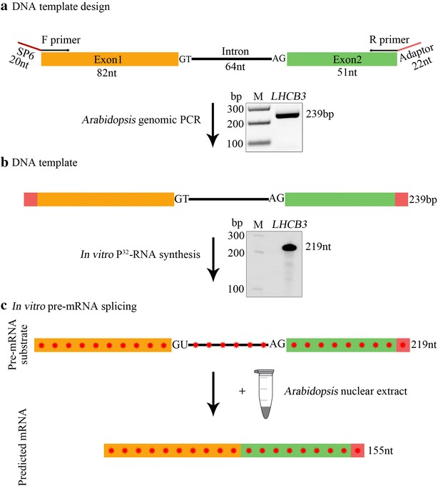
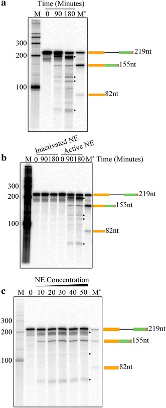
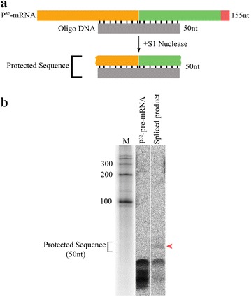
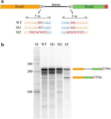
Results: Here we report an in vitro splicing assay system using plant nuclear extract. Several lines of evidence indicate that nuclear extract derived from Arabidopsis seedlings can convert pre-mRNA substrate (LHCB3) into a spliced product. These include: (1) generation of an RNA product that corresponds to the size of expected mRNA, (2) a junction-mapping assay using S1 nuclease revealed that the two exons are spliced together, (3) the reaction conditions are similar to those found with non-plant extracts and (4) finally mutations in conserved donor and acceptor sites abolished the production of the spliced product.

Conclusions: This first report on the plant in vitro splicing assay opens new avenues to investigate plant spliceosome assembly and composition, and splicing regulatory mechanisms specific to plants.

Keywords: Arabidopsis; In vitro splicing; Plant in vitro splicing; Pre-mRNA; Splicing.


Figures

Fig. 1
Fig. 1
Preparation of LHCB3 [32P]-labeled pre-mRNA substrate used for in vitro splicing assay. a Top, a schematic representation of a region of Arabidopsis LHCB3 (AT5G54270) gene used to prepare DNA template to synthesize pre-mRNA substrate. A portion of the third and fourth exons (Orange and green boxes, respectively, labeled as exon 1 and exon2) and second intron (black line, labeled as intron) was used. F primer, forward primer with SP6 promoter sequence (red line), R primer reverse primer with an adaptor sequence (red line). Bottom, PCR fragment amplified with F and R primers using Arabidopsis genomic DNA. The PCR product was gel purified and used for in vitro transcription reaction. b Top, schematic of DNA template that was used to synthesize [32P]-labeled LHCB3 pre-mRNA substrate. Bottom, A representative autoradiogram of in vitro [32P]-labeled LHCB3 pre-mRNA substrate. c Description of in vitro splicing assay. Top, Schematic representation of labeled LHCB3 pre-mRNA substrate used for in vitro splicing assay. Bottom, predicted mRNA after in vitro splicing of pre-mRNA substrate. Sizes of intron, exons, pre-mRNA, and predicted mRNA are indicated. Red asterisks indicate [32P]-nucleotides in RNA
Fig. 2
Fig. 2
In vitro splicing assays. a In vitro splicing assay with the Arabidopsis LHCB3 pre-mRNA substrate. Radioactive LHCB3 pre-mRNA substrate was synthesized in vitro with a DNA template using SP6 RNA polymerase (see Fig. 1b) as described in materials and methods. [32P]-labeled LHCB3 pre-mRNA substrate (25,000 cpm) was incubated with nuclear extract from Arabidopsis etiolated seedlings at 30 °C as described in materials and methods. Samples were withdrawn at intervals (0, 90 and 180 min), [32P]-RNA was extracted and analyzed by electrophoresis on a 6% polyacrylamide gel containing 7 M urea. The gel was dried and exposed to a phosphor-imaging screen. b Heat-inactivation of Arabidopsis NE abolished the production of a spliced product. NE from Arabidopsis etiolated seedlings was incubated at 90 °C for 3 min or kept on ice (as a control) were used for splicing assays at 30 °C with the LHCB3 [32P]-pre-mRNA. Samples were withdrawn at different time points (0, 90, and 180 min), [32P]-RNA was extracted and analyzed as described above. c The spliced product is increased with increasing nuclear extract concentration. In vitro splicing of [32P]-labeled LHCB3 pre-mRNA substrate (25,000 cpm) was carried out at 30 °C in 25 μl reaction volume containing different concentrations 0–50% (v/v) of nuclear extract as described in materials and methods. All reactions were stopped after three hours; [32P]-RNA was extracted and analyzed as described above. M indicates [32P]-labeled RNA markers synthesized in vitro using RNA Century™-Plus Marker Templates (Applied Biosystems, AM7782). M* lane contains [32P]-labeled LHCB3 pre-mRNA, spliced mRNA, and exon1. Schematic diagrams on the right show pre-mRNA, spliced mRNA and exon 1 and their sizes. One of the [32P]-RNA products formed in in vitro splicing assay corresponds to the size of spliced [32P]-mRNA marker, suggesting that it could be a spliced product. The asterisks indicate the potential splicing intermediates. Other [32P]-RNA products could be another pre-mRNA splicing intermediates and/or degradation products
Fig. 3
Fig. 3
Characterization of the spliced product using S1 nuclease. a Schematic representation of S1 nuclease protection assay. Top, a diagram of the hybrid formed between spliced RNA and DNA oligonucleotide (50 nt) complementary to exons junction. Bottom, A diagram of protected sequences after S1 nuclease digestion. b Spliced [32P]-RNA produced in in vitro splicing assay was gel purified as described in Materials and methods. [32P]-pre-mRNA (negative control) and spliced product [32P]-RNA were hybridized to oligo DNA that is complementary to exons junction. Following hybridization, [32P]-RNA–DNA hybrids were digested with S1 nuclease that degraded single stranded nucleic acids. The size of the protected region (50 nts) is indicated. Red arrowhead shows protected exon junction sequence in the spliced product
Fig. 4
Fig. 4
Mutations in conserved splice sites of LHCB3 pre-mRNA substrate modulated the production of the spliced product. a The diagram shows sequence substitutions of conserved 5′ GU and 3′ AG splice sites (ss) of LHCB3 pre-mRNA substrate. DNA templates with different splice site mutations (M1 and M2) were synthesized (Integrated DNA Technologies, Inc., Coralville, IA, USA) for in vitro [32P]-labelled RNA synthesis. b In vitro splicing of [32P]-labeled wild type and two mutants (M1 and M2) of LHCB3 pre-mRNA substrate was carried as described before. Reactions were stopped after 3 h. [32P]-RNA was isolated and analyzed by electrophoresis as above. RNA markers (M and M*) and schematic diagrams on the right were described in Fig. 2 legend
Fig. 5
Fig. 5
Analysis of optimum conditions for splicing assay. a The amount of spliced product at different temperatures. In vitro splicing of LHCB3 [32P]-pre-mRNA substrate (25,000 cpm) was carried out as described earlier at different temperatures (24, 30, 37, and 40 °C). b Addition of ATP to in vitro splicing assay increased the amount of spliced product from LHCB3 pre-mRNA. c Effect of various concentrations of Mg2+ on the production of the spliced product. In vitro splicing reaction of LHCB3 [32P]-pre-mRNA substrate (25,000 cpm) was performed as described previously with different concentrations of Mg2+ (2.5 and 5 mM), or in the presence of different concentration (2,5 and 5 mM) of EDTA, a divalent cation chelator (EDTA). In vitro splicing reaction of LHCB3 [32P]-pre-mRNA substrate (25,000 cpm) was carried out as described above without (0 mM) with increasing concentrations ATP (1, 2 and 3 mM). All reactions were stopped after 3 h. [32P]-RNA was recovered and analyzed by electrophoresis on a 6% polyacrylamide-7 M urea gel, followed by autoradiography. RNA markers (M and M*) and schematic diagrams on the right were described in Fig. 2. The asterisks indicate the potential splicing intermediates. Other [32P]-RNA products could be another pre-mRNA splicing intermediates and/or degradation products

Similar articles

Cited by

References

    1. Hernandez N, Keller W. Splicing of in vitro synthesized messenger RNA precursors in HeLa cell extracts. Cell. 1983;35(1):89–99. - PubMed
    1. Krainer AR, Maniatis T, Ruskin B, Green MR. Normal and mutant human beta-globin pre-mRNAs are faithfully and efficiently spliced in vitro. Cell. 1984;36(4):993–1005. - PubMed
    1. Padgett RA, Konarska MM, Grabowski PJ, Hardy SF, Sharp PA. Lariat RNA’s as intermediates and products in the splicing of messenger RNA precursors. Science. 1984;225(4665):898–903. - PubMed
    1. Lin RJ, Newman AJ, Cheng SC, Abelson J. Yeast mRNA splicing in vitro. J Biol Chem. 1985;260(27):14780–14792. - PubMed
    1. Rio DC. Accurate and efficient pre-mRNA splicing in Drosophila cell-free extracts. Proc Natl Acad Sci USA. 1988;85(9):2904–2908. - PMC - PubMed

LinkOut - more resources