

Teratoma as unusual cause of chest pain, hemoptysis and dyspnea in a young patient
- PMID: 29321967
- PMCID: PMC5752334
- DOI: 10.1016/j.rmcr.2017.12.006
Teratoma as unusual cause of chest pain, hemoptysis and dyspnea in a young patient
Abstract
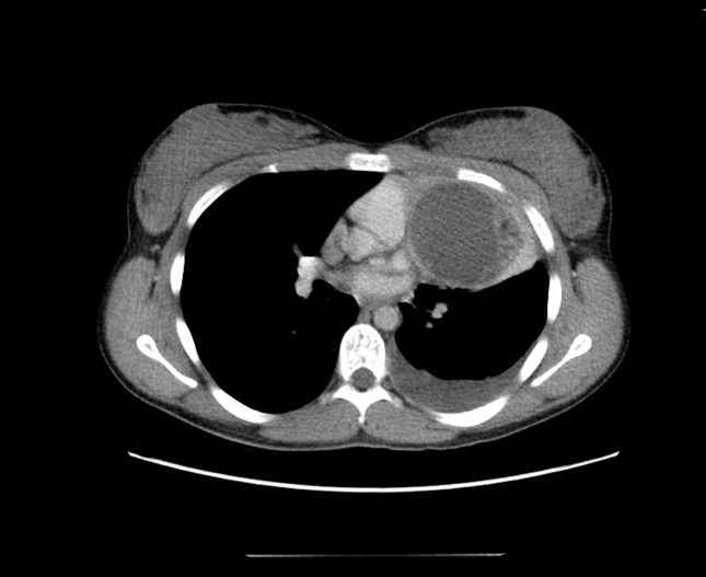
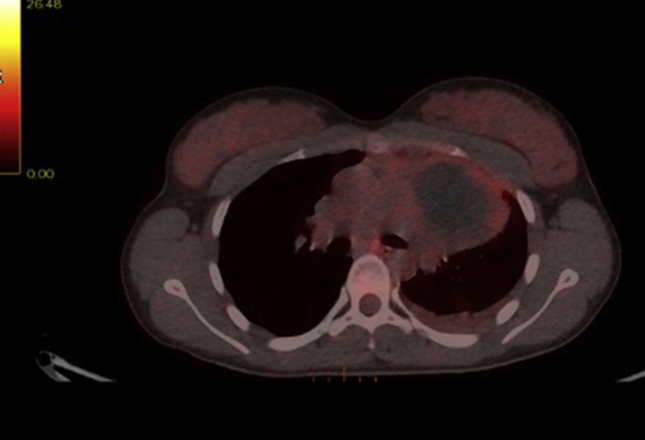
A 16-year-old girl presented with intermittent left chest pain and breathlessness on exertion for last 4 months with one episode of haemoptysis. There has been loss of appetite and weight loss of 4 kg over a period of 1 month. A chest radiograph revealed a large mass like opacity with pleural effusion in the left lung field. Computerized Tomography scanning (CT scanning) and Positron Emission Tomography/Computerized Tomography scanning (PET/CT scanning) demonstrated a 7 cm round, cystic lesion in the anterior mediastinum. Pleural fluid cytology did not show any malignant cell. The patient was referred to cardiothoracic department for thoracotomy and resection. Surgery was uncomplicated with rapid recovery. Histologic findings suggested mature teratoma components surrounded by oedematous pleura and pericardium with adjacent thymus and lung tissue.
Figures

References
-
- Mulen B., Richardson J.D. Primary anterior mediastinal tumors in children and adults. Ann. Thorac. Surg. 1986;42:338. - PubMed
Publication types
LinkOut - more resources
Full Text Sources
Other Literature Sources
