Review
. 2017 Dec 13:5:109.
doi: 10.3389/fcell.2017.00109. eCollection 2017.

Multiple Duties for Spindle Assembly Checkpoint Kinases in Meiosis

Affiliations
Review

Multiple Duties for Spindle Assembly Checkpoint Kinases in Meiosis

Adele L Marston et al. Front Cell Dev Biol. .

Abstract

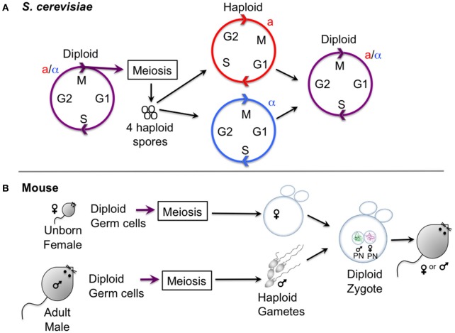
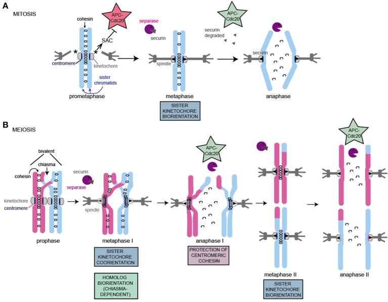
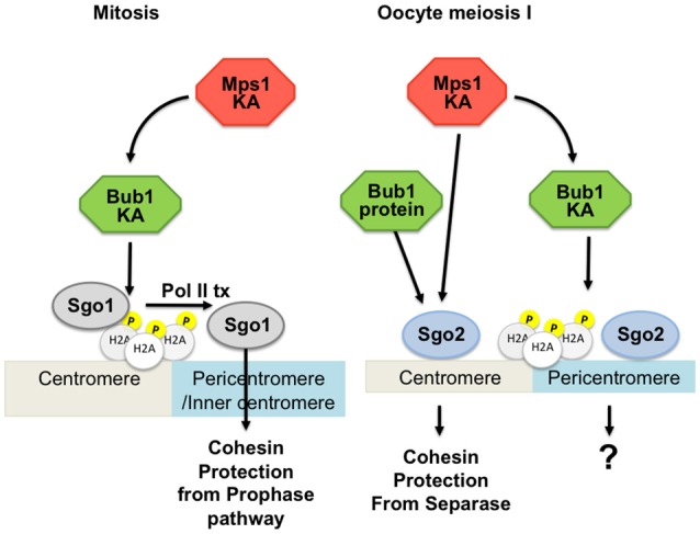
Cell division in mitosis and meiosis is governed by evolutionary highly conserved protein kinases and phosphatases, controlling the timely execution of key events such as nuclear envelope breakdown, spindle assembly, chromosome attachment to the spindle and chromosome segregation, and cell cycle exit. In mitosis, the spindle assembly checkpoint (SAC) controls the proper attachment to and alignment of chromosomes on the spindle. The SAC detects errors and induces a cell cycle arrest in metaphase, preventing chromatid separation. Once all chromosomes are properly attached, the SAC-dependent arrest is relieved and chromatids separate evenly into daughter cells. The signaling cascade leading to checkpoint arrest depends on several protein kinases that are conserved from yeast to man. In meiosis, haploid cells containing new genetic combinations are generated from a diploid cell through two specialized cell divisions. Though apparently less robust, SAC control also exists in meiosis. Recently, it has emerged that SAC kinases have additional roles in executing accurate chromosome segregation during the meiotic divisions. Here, we summarize the main differences between mitotic and meiotic cell divisions, and explain why meiotic divisions pose special challenges for correct chromosome segregation. The less-known meiotic roles of the SAC kinases are described, with a focus on two model systems: yeast and mouse oocytes. The meiotic roles of the canonical checkpoint kinases Bub1, Mps1, the pseudokinase BubR1 (Mad3), and Aurora B and C (Ipl1) will be discussed. Insights into the molecular signaling pathways that bring about the special chromosome segregation pattern during meiosis will help us understand why human oocytes are so frequently aneuploid.

Keywords: Aurora B-like kinases; Bub1; BubR1/Mad3; Mps1; chromosome congression; cohesin protection; meiosis; spindle assembly checkpoint.


Figures

Figure 1
Figure 1
Gametogenesis in yeast and female mice. (A) Outline of the lifecycle of the budding yeast S. cerevisiae, which can propagate vegetatively through mitotic division in both its haploid and diploid form. Haploid yeast of opposite mating types (a and alpha) mate and undergo conjugation to generate a zygote, upon which nuclei fuse (karyogamy). Starvation of diploid cells triggers meiosis, culminating in the production of four haploid spores. Note that the lifestyle of S. pombe is similar, except that meiosis and sporulation occur directly after karyogamy so that diploid cells are short-lived. (B) Oocyte differentiation in female mice. Gametes are generated from diploid germ cells, in the female embryo before birth for oocytes, and in the adult male for spermatocytes. After hormonal stimulation in adult females, some oocytes that are arrested in prophase I undergo a growth phase. During the menstrual cycle usually one (humans) or several (mouse) oocytes are induced to undergo meiosis I and enter meiosis II, where they will remain arrested to await fertilization with a male gamete that has already finished meiosis I and II and has progressed into G1 phase. Oocytes exit meiosis II after fertilization, and the male and female pronucleus fuse to form the diploid zygote, the first cell of the embryo.
Figure 2
Figure 2
Schematic of chromosome segregation during mitosis and meiosis. The key adaptations to meiotic chromosomes are indicated. Note that while a single microtubule contacts each kinetochore (mitosis, meiosis II) or pair of kinetochores (meiosis I) in S. cerevisiae in most organisms, multiple microtubules connect to each kinetochore, resulting in increased probability and configurations of incorrect attachments in both mitosis and meiosis. (A) Key features of mitotic chromosome segregation. (B) Key features and adaptations of meiotic chromosome segregation. For details see text.
Figure 3
Figure 3
Involvement of Bub1 and Mps1 kinases in Sgo2 localization in mouse oocytes. On the left: Sgo1 in mitosis is localized by Bub1-dependent phosphorylation of H2A and transcription for protection of cohesin. On the right: Two pools of Sgo2 can be distinguished in oocyte meiosis I, one of which is localized independently of H2A phosphorylation by Bub1, and which is required for protection of centromeric cohesin. Mps1 kinase is involved in localizing the pool of Sgo2 required for protection, at the centromere. Bub1 kinase is required for localizing a pool of Sgo2 in an H2A-phosphorylation dependent manner, but this pool is largely dispensible for protection of centromeric cohesin in meiosis I. KA, kinase activity; Pol II tx, Polymerase II dependent transcription.

References

    1. Argüello-Miranda O., Zagoriy I., Mengoli V., Rojas J., Jonak K., Oz T., et al. (2017). Casein kinase 1 coordinates cohesin cleavage, gametogenesis, and exit from M phase in meiosis II. Dev. Cell 40, 37–52. 10.1016/j.devcel.2016.11.021 - DOI - PubMed
    1. Asghar A., Lajeunesse A., Dulla K., Combes G., Thebault P., Nigg E. A., et al. (2015). Bub1 autophosphorylation feeds back to regulate kinetochore docking and promote localized substrate phosphorylation. Nat. Commun. 6:8364. 10.1038/ncomms9364 - DOI - PMC - PubMed
    1. Bähler J., Schuchert P., Grimm C., Kohli J. (1991). Synchronized meiosis and recombination in fission yeast: observations with pat1-114 diploid cells. Curr. Genet. 19, 445–451. 10.1007/BF00312735 - DOI - PubMed
    1. Balboula A. Z., Schindler K. (2014). Selective disruption of aurora C kinase reveals distinct functions from aurora B kinase during meiosis in mouse oocytes. PLoS Genet. 10:e1004194. 10.1371/journal.pgen.1004194 - DOI - PMC - PubMed
    1. Batiha O., Swan A. (2012). Evidence that the spindle assembly checkpoint does not regulate APC(Fzy) activity in Drosophila female meiosis. Genome 55, 63–67. 10.1139/g11-079 - DOI - PubMed

LinkOut - more resources