. 2018 Sep;80(3):864-873.
doi: 10.1002/mrm.27077. Epub 2018 Jan 10.

Development of methods and feasibility of using hyperpolarized carbon-13 imaging data for evaluating brain metabolism in patient studies

Affiliations

Development of methods and feasibility of using hyperpolarized carbon-13 imaging data for evaluating brain metabolism in patient studies

Ilwoo Park et al. Magn Reson Med. 2018 Sep.

Abstract

Purpose: Hyperpolarized 13C metabolic imaging is a non-invasive imaging modality for evaluating real-time metabolism. The purpose of this study was to develop and implement experimental strategies for using [1-13C]pyruvate to probe in vivo metabolism for patients with brain tumors and other neurological diseases.

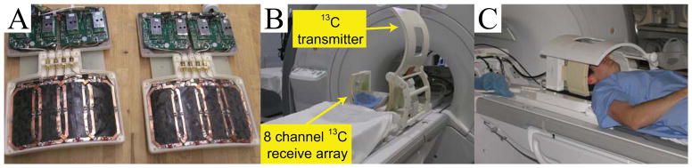
Methods: The 13C RF coils and pulse sequences were tested in a phantom and were performed using a 3T whole body scanner. Samples of [1-13C]pyruvate were polarized using a SPINlab system. Dynamic 13C data were acquired from eight patients previously diagnosed with brain tumors, who had received treatment and were being followed with serial MR scans.

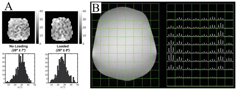
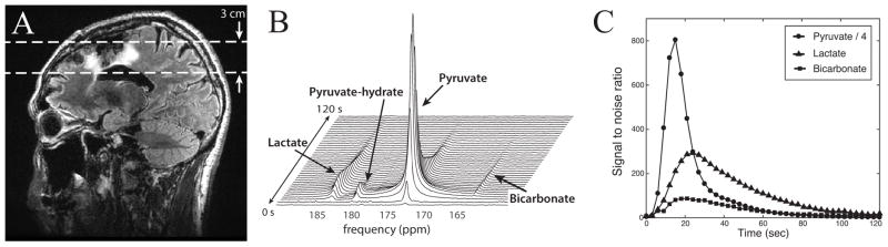
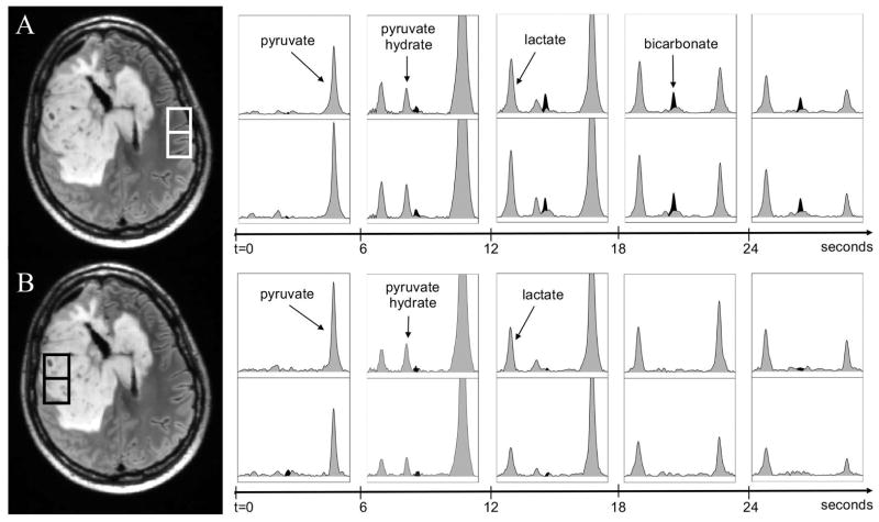
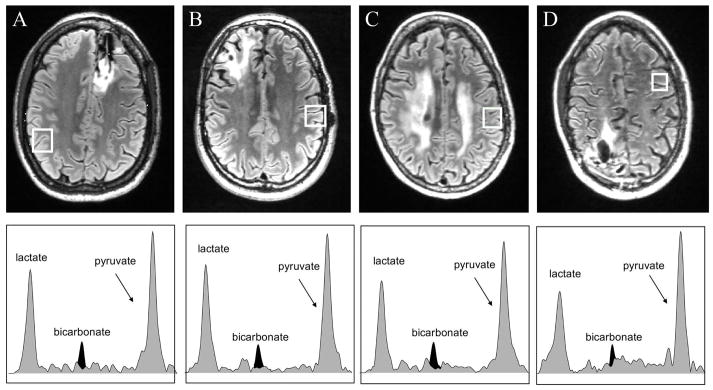
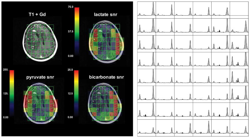
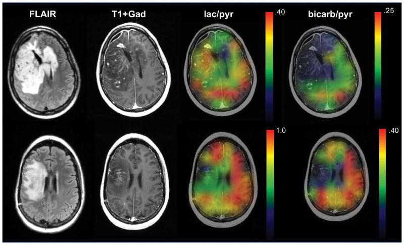
Results: The phantom studies produced good quality spectra with a reduction in signal intensity in the center due to the reception profiles of the 13C receive coils. Dynamic data obtained from a 3 cm slice through a patient’s brain following injection with [1-13C]pyruvate showed the anticipated arrival of the agent, its conversion to lactate and bicarbonate, and subsequent reduction in signal intensity. A similar temporal pattern was observed in 2D dynamic patient studies, with signals corresponding to pyruvate, lactate and bicarbonate being in normal appearing brain but only pyruvate and lactate being detected in regions corresponding to the anatomic lesion. Physiological monitoring and follow-up confirmed that there were no adverse events associated with the injection.

Conclusions: This study has presented the first application of hyperpolarized 13C metabolic imaging in patients with brain tumor and demonstrated the safety and feasibility of using hyperpolarized [1-13C]pyruvate to evaluate in vivo brain metabolism.

Keywords: brain tumor patients; dynamic nuclear polarization; hyperpolarized carbon-13 MRI.


Figures

Figure 1
Figure 1
13C RF coil configurations developed for human brain study. (A) 8-channel 13C phased array coils. (B) Clamshell volumetric 13C transmit coil and bilateral eight-channel phased array receive coils. (C) A picture of 13C RF coil setup with a volunteer.
Figure 2
Figure 2
Results from the 13C coil loading and initial data acquisition tests. (A) B1+ maps of flip angle were calculated using a double flip angle method and showed that the B1 maps were similar between the unloaded and loaded conditions. (B) 2D EPSI data from the head-shaped phantom containing ethylene glycol demonstrated the combined reception profile of the receive coils.
Figure 3
Figure 3
Slice-localized hyperpolarized 13C dynamic data from patient 1. (A) T2 FLAIR image in a sagittal plane shows the hyperintense region around the resection cavity and the ventricle. (B) The stack plot of 13C magnitude spectra, showing a temporal evolution of lactate, pyruvate and bicarbonate signal from the brain. (C) The SNR of lactate, pyruvate, urea and bicarbonate are plotted over time. The pyruvate SNR was divided by 4 so that it could be viewed on the same graph as the pyruvate and bicarbonate.
Figure 4
Figure 4
Temporal changes in spectra from the highlighted voxels in (A) normal appearing brain and (B) the T2 lesion from patient 2. The spectra with peaks shaded in gray are from the data reconstructed at the acquired reference frequency and the peaks shaded in black represent the data reconstructed at the bicarbonate frequency. The spectra shown are from time at 0s, 6s, 12s, 18s and 24s from the start of data acquisition. The highest pyruvate and pyruvate-hydrate occur at 6s, while the highest lactate occurs at time point 15s. It can be seen that there are lactate and bicarbonate peaks in normal appearing brain and lactate peaks in the T2 lesion during the period of 9s to 24s. The chemical shift range is 186.46 to 169.61ppm for all spectroscopic voxels. The intensity scales are arbitrary, but were kept the same for all spectra.
Figure 5
Figure 5
FLAIR images and spectra in the highlighted normal appearing brain voxels at time with maximum lactate from (A) patient 3, (B) patient 4, (C) patient 5 and (D) patient 6. The spectra with peaks shaded in gray are from the data reconstructed at the acquired reference frequency and the peaks shaded in black represent the data reconstructed at the bicarbonate frequency. The relative levels of pyruvate are similar but the lactate is slightly lower in all patients shown and the bicarbonate is not detectable for patient 6, for whom the acquisition had the smallest voxel size 1.5x1.5x2 cm and 12 rather than 10 phase encodes. The chemical shift range is 186.46 to 169.61ppm for all spectroscopic voxels. The intensity scales are arbitrary, and were manually set to have similar pyruvate intensity levels between subjects.
Figure 6
Figure 6
A post-gadolinium T1-weighted image, color maps of integrated pyruvate, lactate and bicarbonate SNR and arrays of summed spectra from patients 2. The spectra with peaks shaded in gray are from the data reconstructed at the acquired reference frequency and the peaks shaded in black represent the data reconstructed at the bicarbonate frequency. The lactate and pyruvate peak intensities were determined from the former gray spectra and the bicarbonate peak intensities from the black spectra. The proposed coil setup with the clamshell transmit and the 8-channel bilateral receive arrays allowed the acquisition of 13C signals across the majority of the brain but with significantly lower SNR in central regions. The low SNR in the center of the brain was accentuated by reduced signal in the ventricles.
Figure 7
Figure 7
Anatomic and interpolated hyperpolarized 13C metabolite ratio image overlays estimated from 2D EPSI dynamic data acquired at 2 cm in plane resolution and 2 cm slice thickness from 2 patients with treated GBM. The differences in scales for the color overlay images are due to the single-band constant flip angle excitation scheme employed for patient 2 and the multi-band variable flip angle excitation scheme used for patient 8. Note that in both cases the bicarbonate/pyruvate was higher in regions of normal appearing brain. The patient in the upper images (patient 2) had progressive tumor at the time of this exam and the patient in the lower images (patient 8) was not characterizing as progressing until subsequent follow-up scans. lac/pyr and bicarb/pyr represent lactate/pyruvate and bicarbonate/pyruvate, respectively.

References

    1. Ardenkjaer-Larsen JH, Fridlund B, Gram A, Hansson G, Hansson L, Lerche MH, et al. Increase in signal-to-noise ratio of > 10,000 times in liquid-state NMR. Proceedings of the National Academy of Sciences of the United States of America. 2003;100(18):10158–63. - PMC - PubMed
    1. Park I, Hu S, Bok R, Ozawa T, Ito M, Mukherjee J, et al. Evaluation of heterogeneous metabolic profile in an orthotopic human glioblastoma xenograft model using compressed sensing hyperpolarized 3D 13C magnetic resonance spectroscopic imaging. Magnetic resonance in medicine. 2013;70(1):33–9. - PMC - PubMed
    1. Brindle K. New approaches for imaging tumour responses to treatment. Nature reviews Cancer. 2008;8(2):94–107. - PubMed
    1. Warburg O. On the origin of cancer cells. Science (New York, NY) 1956;123(3191):309–14. - PubMed
    1. Albers MJ, Bok R, Chen AP, Cunningham CH, Zierhut ML, Zhang VY, et al. Hyperpolarized 13C lactate, pyruvate, and alanine: noninvasive biomarkers for prostate cancer detection and grading. Cancer research. 2008;68(20):8607–15. - PMC - PubMed

Publication types