. 2018 Jan 11;9(1):164.
doi: 10.1038/s41467-017-02637-3.

Signaling ammonium across membranes through an ammonium sensor histidine kinase

Affiliations

Signaling ammonium across membranes through an ammonium sensor histidine kinase

Tobias Pflüger et al. Nat Commun. .

Abstract

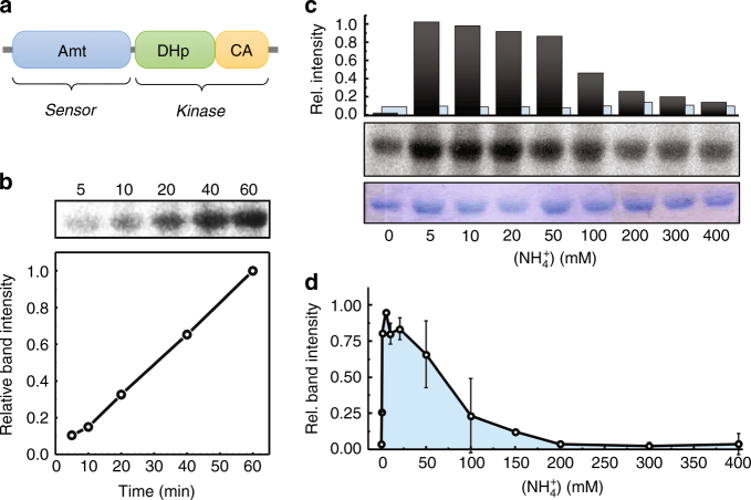
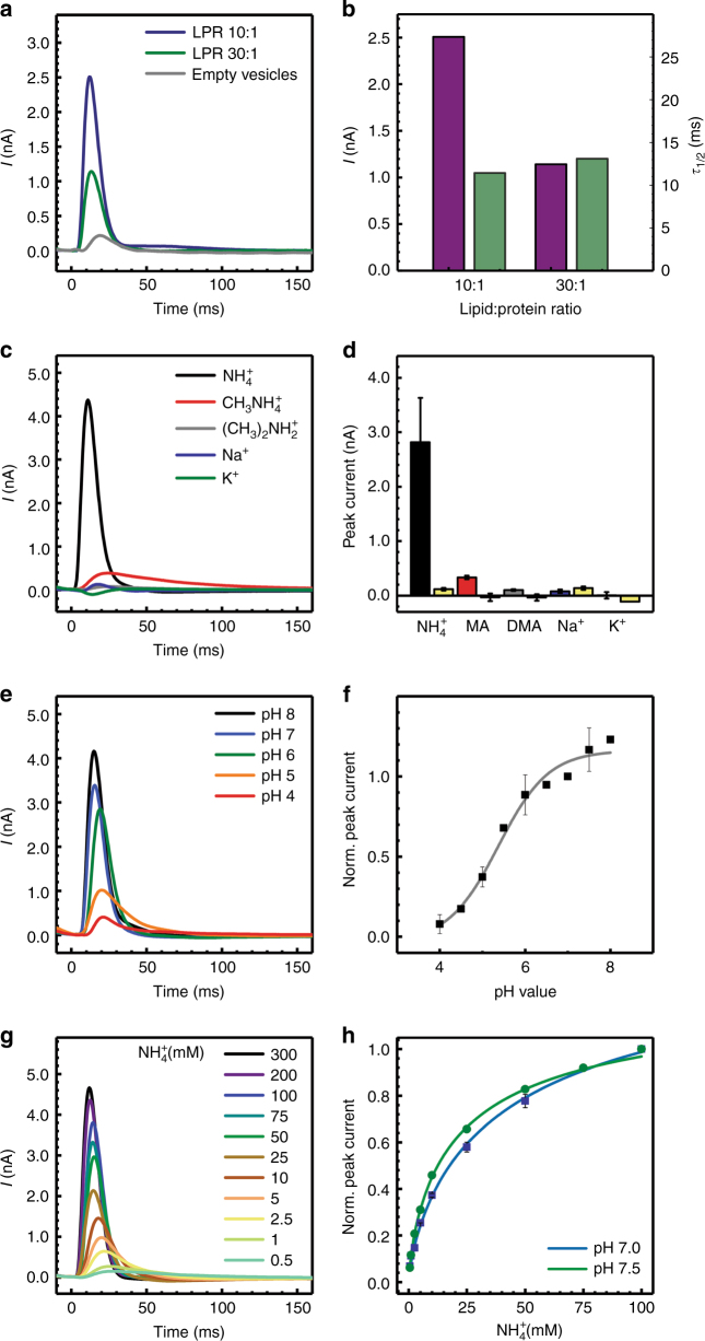
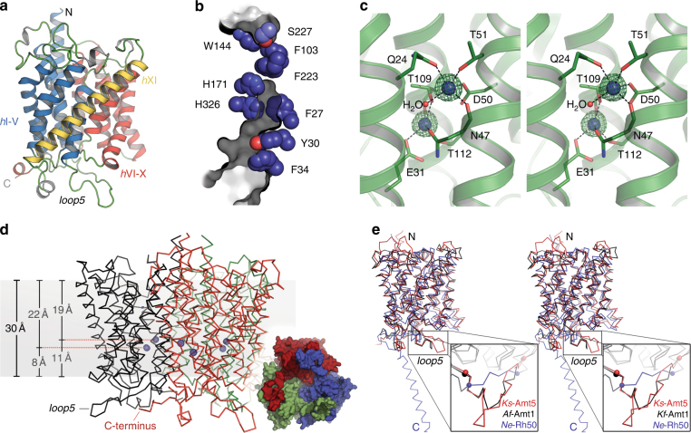
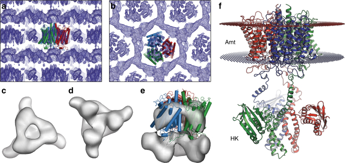
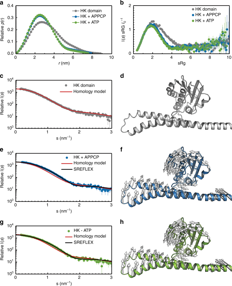
Sensing and uptake of external ammonium is essential for anaerobic ammonium-oxidizing (anammox) bacteria, and is typically the domain of the ubiquitous Amt/Rh ammonium transporters. Here, we report on the structure and function of an ammonium sensor/transducer from the anammox bacterium "Candidatus Kuenenia stuttgartiensis" that combines a membrane-integral ammonium transporter domain with a fused histidine kinase. It contains a high-affinity ammonium binding site not present in assimilatory Amt proteins. The levels of phosphorylated histidine in the kinase are coupled to the presence of ammonium, as conformational changes during signal recognition by the Amt module are transduced internally to modulate the kinase activity. The structural analysis of this ammonium sensor by X-ray crystallography and small-angle X-ray-scattering reveals a flexible, bipartite system that recruits a large uptake transporter as a sensory module and modulates its functionality to achieve a mechanistic coupling to a kinase domain in order to trigger downstream signaling events.


Conflict of interest statement

The authors declare no competing financial interests.

Figures

Fig. 1
Fig. 1
Architecture of Ks-Amt5 and its kinase activity. a The ammonium sensor/transducer Ks-Amt5 comprises a membrane-integral sensor domain (Amt) and a soluble, C-terminally fused histidine kinase, consisting of a DHp and a CA subdomain. b With [γ-32P]-ATP, the isolated HK domain alone shows increasing, but unregulated autophosphorylation over time. c In contrast, only full-length Ks-Amt5 reacts to variations in [NH4+] and shows rapidly increasing phosphorylation levels (black bars) at low [NH4+] that drop again at higher concentrations, whereas protein levels (Coomassie-stained, blue bars) remain constant. All data points were measured after 60 min incubation. d In an average of multiple (n = 5) experiments on a linear time scale, variations in phosphorylation levels of residue H406 show the rapid response of Ks-Amt5 at low [NH4+], followed by a slower decrease towards high concentrations of substrate, where passive diffusion of NH3 can lead to unregulated permeation across the lipid bilayer
Fig. 2
Fig. 2
Electrophysiological analysis of Ks-Amt5 in proteoliposomes. a Transient currents recorded at lipid:protein ratios (LPR) of 10:1 and 30:1 vs. protein-free liposomes reveal specific charge displacement events in the presence of Ks-Amt5. b Bar graph of the peak current (violet, nA) and half-maximum decay time (green, ms) values from a. The unchanged decay time points towards charge displacement rather than ion translocation. c Transient currents recorded at pH 7.5 for different cations (300 mM) at a LPR of 10:1. Ks-Amt5 is highly specific for NH4+, but does not act on methylammonium (MA) or dimethylammonium (DMA), which are alternative substrates of assimilatory Amt proteins. d Bar graph of the peak currents measured in c and the respective backgrounds (yellow). Only NH4+, but not MA or DMA, triggered an electrogenic response. e pH-dependent transients induced by 300 mM NH4+ concentration jumps. f Variation of normalized peak currents with pH (as in e). The fit (solid line) used a titration function yielding a pKapp of 5.5. g Saturation behavior of NH4+ transients at pH 7.5 with increasing concentration of the ion. h Normalized peak currents with respect to NH4+ concentration at pH 7.5 and 7.0. The affinity for the cations is increased at the higher pH value (K0.5app, pH 7.5 = 22.9 ± 0.87 mM fitted with a Hill coefficient of 0.75). All experiments were done at least in triplicate on two different protein reconstitutions and two separate protein batches. Error bars are thus calculated from a minimum of n = 12
Fig. 3
Fig. 3
High-resolution structure of the Amt domain of Ks-Amt5. a The monomer of Ks-Amt5 exhibits the typical transporter fold of Amt/Rh family proteins, with two groups of five transmembrane helices (hI-V, hVI-X) related through a twofold pseudosymmetry axis in the membrane plane, stabilized through the clamping helix hXI. The C-terminal HK domain was undefined due to structural disorder. b The Amt domain retains all functionally relevant residues (orientation as in a), in particular the selectivity filter (W144/S227), the Phe gate (F103, F223) and the His pair (H171, H326). However, in comparison with the Af-Amt1 transporter, for which the transport channel is overlaid in gray, an inward shift of the His pair in combination with three bulkier residues (F27, Y30, F34) in helix hI effectively closes the transmembrane transport pathway for ammonium ions. c Stereo representation of the NH4+ binding sites within the membrane, with an FoFc omit difference electron density map contoured at the 5.0 σ level. See Supplementary Figure 7 for the relative orientation of the ion binding site and the transport channel. d Side view of the trimer, indicating the location of the NH4+ binding sites within the membrane (gray). The inset shows a bottom view of the trimer, highlighting how the C-termini extend over the neighboring monomer towards loop 5. e Superposition of Ks-Amt5 (red) with Af-Amt1 (black) and the Rh orthologue Ne-Rh50 (blue). The latter contains a helical extension at the C-terminus that adopts a different conformation than the connection of the HK domain in Ks-Amt5. Although the three structures are homologous in the membrane-integral part, they differ strongly in length and conformation of the functionally relevant loop 5 (bordered by balls) that connects the symmetry-related halves of each monomer
Fig. 4
Fig. 4
Low-resolution analysis of full-length Ks-Amt5. a Electron density map showing the strata of the type I crystals, with the high-resolution structure of the Amt domain placed and shown in cartoon representation. b Top view of a. The electron density map is better resolved for the Amt domain that determines the crystal packing, whereas the HK domain shows disorder and attains a position between neighboring Amt domains. c Top view of full-length Ks-Amt5 in a weighted electron density synthesis at 8 Å resolution contoured at the 1σ level. d Bottom view onto the HK domain. e Low-resolution electron density synthesis of Ks-Amt5, superimposed with the high-resolution 3D structure of the Amt domain. f Hypothetical, extended arrangement of the sensor-transducer in the membrane, showing the structure of the Amt domain linked to a homology model for the HK domain in an arbitrary, extended conformation. The HK domains are structurally flexible in the cytoplasm, and their conformation in solution is not identical to the one observed in the crystal lattice (Supplementary Figure 10a)
Fig. 5
Fig. 5
SAXS analysis of nucleotide binding to the HK domain. Real-space distance distributions p(r) of the HK domain with no ligand (gray) and in the presence of 10 µM of APPCP (blue), or 10 µM of ATP (green). b Dimensionless Kratky plot of the data in a, highlighting the change toward a more compact and less-flexible structure when either APPCP or ATP are added to the HK domain. c SAXS data for the ligand-free HK domain, fitted with the homology model depicted in d. e SAXS data for the Mg-APPCP-bound HK domain, fitted with the homology model (red), as well as with the best SAXS-refined elastic network conformation (black). f Conformational transitions from the template homology model refined against the Mg-APPCP-bound data in e. Vectors describe the direction of motion. g SAXS data and corresponding fits as in e for the Mg-ATP bound HK domain. h The derived conformational transitions from g are virtually identical to the ones obtained for Mg-APPCP in f. No effect was noted upon addition of the protein kinase C inhibitor Ro 31-8220

References

    1. Jetten MSM, et al. Biochemistry and molecular biology of anammox bacteria. Crit. Rev. Biochem. Mol. Biol. 2009;44:65–84. doi: 10.1080/10409230902722783. - DOI - PubMed
    1. Andrade SLA, Einsle O. The Amt/Mep/Rh family of ammonium transport proteins. Mol. Membr. Biol. 2007;24:357–365. doi: 10.1080/09687680701388423. - DOI - PubMed
    1. Kartal B, et al. Molecular mechanism of anaerobic ammonium oxidation. Nature. 2011;479:127–130. doi: 10.1038/nature10453. - DOI - PubMed
    1. Willett JW, Kirby JR. Genetic and biochemical dissection of a HisKA domain identifies residues required exclusively for kinase and phosphatase activities. PLoS. Genet. 2012;8:e1003084. doi: 10.1371/journal.pgen.1003084. - DOI - PMC - PubMed
    1. Tremblay PL, Hallenbeck PC. Of blood, brains and bacteria, the Amt/Rh transporter family: emerging role of Amt as a unique microbial sensor. Mol. Microbiol. 2009;71:12–22. doi: 10.1111/j.1365-2958.2008.06514.x. - DOI - PubMed

Publication types

MeSH terms

LinkOut - more resources