. 2018 Jan 11;8(1):461.
doi: 10.1038/s41598-017-17634-1.

Issues with the Specificity of Immunological Reagents for NLRP3: Implications for Age-related Macular Degeneration

Affiliations

Issues with the Specificity of Immunological Reagents for NLRP3: Implications for Age-related Macular Degeneration

Cassandra Kosmidou et al. Sci Rep. .

Abstract

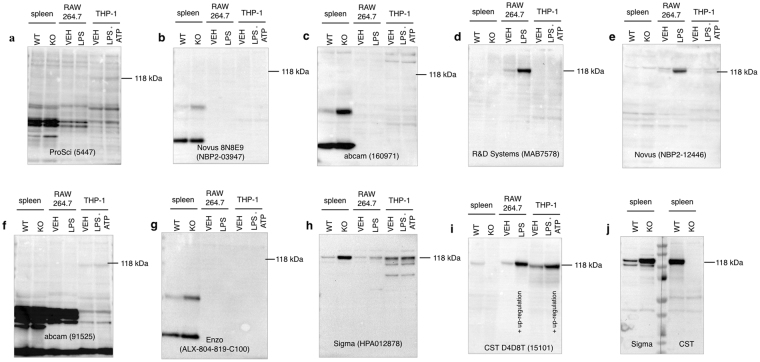
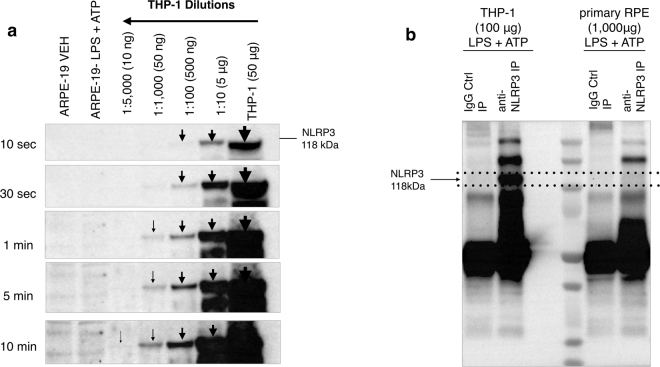
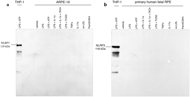
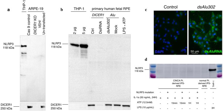
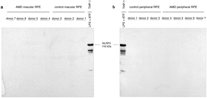
Contradictory data have been presented regarding the implication of the NACHT, LRR and PYD domains-containing protein 3 (NLRP3) inflammasome in age-related macular degeneration (AMD), the leading cause of vision loss in the Western world. Recognizing that antibody specificity may explain this discrepancy and in line with recent National Institutes of Health (NIH) guidelines requiring authentication of key biological resources, the specificity of anti-NLRP3 antibodies was assessed to elucidate whether non-immune RPE cells express NLRP3. Using validated resources, NLRP3 was not detected in human primary or human established RPE cell lines under multiple inflammasome-priming conditions, including purported NLRP3 stimuli in RPE such as DICER1 deletion and Alu RNA transfection. Furthermore, NLRP3 was below detection limits in ex vivo macular RPE from AMD patients, as well as in human induced pluripotent stem cell (hiPSC)-derived RPE from patients with overactive NLRP3 syndrome (Chronic infantile neurologic cutaneous and articulate, CINCA syndrome). Evidence presented in this study provides new data regarding the interpretation of published results reporting NLRP3 expression and upregulation in RPE and addresses the role that this inflammasome plays in AMD pathogenesis.


Conflict of interest statement

The authors declare that they have no competing interests.

Figures

Figure 1
Figure 1
Validating the specificity of commercially available anti-NLRP3 antibodies by Western blotting. Nine commercially available anti-NLRP3 antibodies were tested in terms of their specificity against murine and human positive controls, including mouse spleen tissue, murine RAW 264.7 and human THP-1 macrophage cell lines. Antibody specificity was validated by testing protein expression in spleen tissue from Nlrp3 knockout mice as a negative control. RAW 264.7 cells were primed with (LPS 10 ng/mL) for 6 hours and were compared to vehicle untreated cells. THP-1 macrophages were primed with LPS (10 μg/mL) plus ATP (5 mM) for 3 hours and compared to vehicle control THP-1. 50 μg of total protein were loaded on a gel and blotted with anti-NLRP3 antibodies with the expected molecular weight at ~118 kDa. The positive control panel was blotted with the following anti-NLRP3 antibodies. (a) ProSci, 5447, (b) Novus 8N8E9, NBP2-03947, (c) abcam, 160971, (d) R&D Systems, MAB7578, (e) Novus NBP2-12446, (f) abcam, 91525, (g) Enzo ALX-804-819-C100, (h) Sigma HPA012878 and (i) Cell Signaling Technologies (D4D8T, 15101). Blots were exposed for 10 minutes. CST (15101) shows a specific band at the expected molecular weight for all positive controls, which is absent in spleen from Nlrp3 −/− sample. The response was amplified in stimulated RAW 264.7 and stimulated THP-1 cells compared to vehicle controls, demonstrating an upregulation of NLRP3 protein levels following inflammasome priming. In (j) duplicate samples of wild type and Nlrp3 −/− spleens were run in parallel, membrane was cut to probe with CST (15101) or Sigma (HPA012878) antibodies to verify their differences. An informative list of the antibodies is shown in Supplementary Table S1. Each blot was repeated in three biologically independent replicates (n = 3) and equal protein loading was confirmed by Coomassie Blue staining (Supplementary Fig. S2). WT: wild type, KO: knockout, VEH: vehicle.
Figure 2
Figure 2
Sensitivity and immunoprecipitation assay for anti-NLRP3 antibody CST (D4D8T, 15101). (a) Serial dilutions of THP-1 cell lysate with starting total protein concentration of 50 μg (1:10, 1:100, 1:1000, 1:5000 in lysis buffer) demonstrate that the anti-NLRP3 antibody (CST D4D8T, 15101) was sensitive enough to detect NLRP3 from a protein sample diluted down to 10 ng of total THP-1 protein content. NLRP3 was not detectable in 25 μg protein samples of baseline or stimulated (LPS 10 μg/mL plus ATP 5 Mm, 24 hours) ARPE-19 cells at a 10-minute exposure time. In Fig. 2a the blot is presented at different exposure times (10 sec, 30 sec, 1 min, 5 min, 10 min). In addition, the blot is cropped, thus the full-length blot is presented in Supplementary Figure S7. (b) Protein A/G agarose beads coupled with anti-NLRP3 antibody (CST, 15101) were used to immunoprecipitate NLRP3. An isotype anti-Rabbit IgG antibody was used as a negative control instead of the NLRP3 antibody. NLRP3 protein was successfully immunoprecipitated in vehicle control THP-1 cell lysate but not in primary RPE cells stimulated with LPS (10 μg/mL) and ATP (5 mM) for 24 hours. Equal protein loading was confirmed by Coomassie Blue staining (Supplementary Figure S3).
Figure 3
Figure 3
Absence of NLRP3 expression in human ARPE-19 cell line and primary human fetal RPE under basal and NLRP3-inducing stimuli. NLPR3 protein expression levels were assessed in human ARPE-19 (a) and primary human fetal RPE (b) cell lysates under basal conditions and NLRP3-priming stimuli. Both ARPE-19 and primary RPE cells were treated with LPS (10 μg/mL) alone, LPS (10 μg/mL) plus ATP (5 mM), LPS (10 μg/mL) plus TCDD (10 nM), LPS (20 μg/mL) plus IL-1α (25 ng/mL), LPS (20 μg/mL) plus IL-1α (25 ng/mL) plus 7-Ketocholesterol (10 μM), TNFα (10 ng/mL), IL-17α (100 ng/mL), Pam3CSK4 (300 ng/mL) for 24 hours and oxidized-LDL (500 μg/mL) for 48 hours. NLRP3 expression levels in ARPE-19 and primary RPE cells were compared to stimulated human THP-1 cell lysate LPS (10 μg/mL) plus ATP (5 mM) for 3 hours. The membrane was blotted with anti-NLRP3 antibody (CST, 15101) and no apparent bands were present to indicate NLRP3 expression in either cell line or primary cells at a 10-minute exposure time. Each blot was repeated in three biologically independent replicates (n = 3) and equal protein loading was confirmed by Coomassie Blue staining (Supplementary Figure S4).
Figure 4
Figure 4
Absence of NLRP3 expression in human ARPE-19 cell line and primary human fetal RPE following DICER1 deletion or downregulation and dsAlu RNA transfection. Absence of NLRP3 expression in iPSC-RPE from CINCA patients. NLPR3 protein expression levels were assessed in human ARPE-19 cells following DICER1 knockout via CRISPR/Cas9 (a) and DICER1 knockdown via DsiRNA in primary human fetal RPE (b). Analysis was performed by Western blotting, using the CST NLRP3 antibody (CST, 15101). No observed changes in NLRP3 expression were seen following DICER1 knockout or knockdown compared to Cas9 and mock transfection controls. Transfection of stimulated primary human fetal RPE cells (LPS 10 μg/mL plus ATP 5 mM for 24 hours) with dsAlu302 RNA did not cause any changes in NLRP3 levels compared to mock transfection control (b). Signals were compared to stimulated THP-1 cells LPS (10 μg/mL) plus ATP (5 mM) for 3 hours and images were taken after a 10-minute exposure. Successful transfection of primary RPE cells with dsAlu302 was validated by immunofluorescence compared to control cells (blue: DAPI, green: dsAlu302 RNA) (c). Figure 4d illustrates the absence of NLRP3 protein in differentiated RPE generated from iPSCs from CINCA patients, with and without the NLRP3 mutation. NLRP3 was absent both in baseline and stimulated IL-1α (50 ng/mL, 24 hours), ATP (12.5 mM) CINCA-derived iPSC-RPE cells. Equal Protein loading was confirmed by Coomassie Blue staining (Supplementary Figure S5).
Figure 5
Figure 5
Western blot analysis of NLRP3 in human macular and peripheral RPE from AMD donors and age-matched controls. Human macular and peripheral RPE lysates were collected from four AMD donors (7 globes) and three age-matched non-AMD controls (6 globes). NLRP3 levels in macular and peripheral RPE samples were examined by Western blotting using anti-NLRP3 antibody (CST, 15101). Positive control used included primed THP-1 macrophages (LPS 10 μg/mL and ATP 5 mM, 3 hours). Blots were exposed for 10 minutes and the absence of specific bands in either AMD or age-matched control RPE samples indicates that NLRP3 is unlikely to be expressed in human RPE cells. Protein loading was confirmed by Coomassie Blue staining (Supplementary Figure S6).

References

    1. World Health Organization. Prevention of Blindness and Visual Impairmenthttp://www.who.int/blindness/causes/priority/en/ (2017).
    1. Wong WL, et al. Global prevalence of age-related macular degeneration and disease burden projection for 2020 and 2040: a systematic review and meta-analysis. Lancet Glob Health. 2014;2:e106–116. doi: 10.1016/S2214-109X(13)70145-1. - DOI - PubMed
    1. Miller JW. VEGF: From Discovery to Therapy: The Champalimaud Award Lecture. Transl Vis Sci Technol. 2016;5:9. doi: 10.1167/tvst.5.2.9. - DOI - PMC - PubMed
    1. Rosenfeld PJ, et al. Ranibizumab for Neovascular Age-Related Macular Degeneration. N Eng J Med. 2006;355:1419–1431. doi: 10.1056/NEJMoa054481. - DOI - PubMed
    1. Deangelis MM, Silveira AC, Carr EA, Kim IK. Genetics of age-related macular degeneration: current concepts, future directions. Semin Ophthalmol. 2011;26:77–93. doi: 10.3109/08820538.2011.577129. - DOI - PMC - PubMed

Publication types

MeSH terms