. 2018 Jan 11;8(1):429.
doi: 10.1038/s41598-017-18888-5.

Green tea extract attenuates LPS-induced retinal inflammation in rats

Affiliations

Green tea extract attenuates LPS-induced retinal inflammation in rats

Jia Lin Ren et al. Sci Rep. .

Abstract

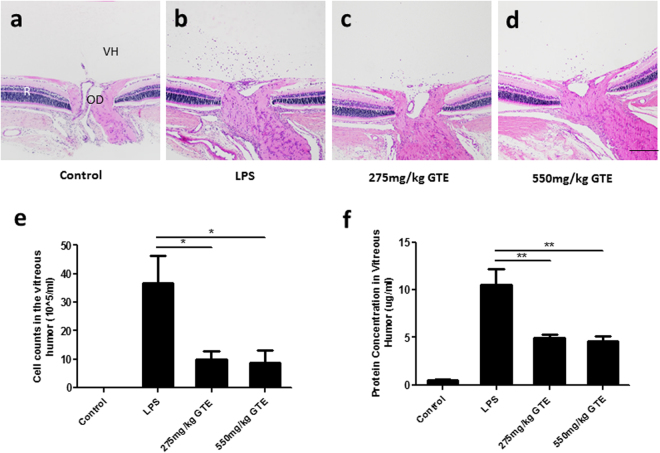
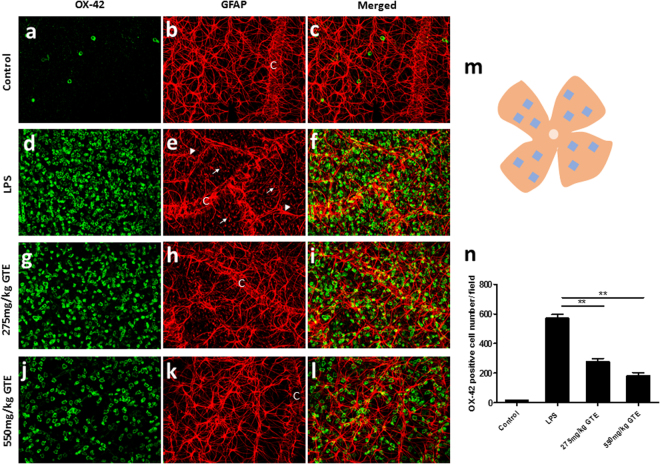
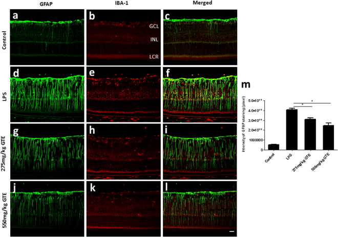
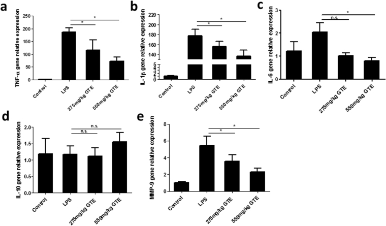
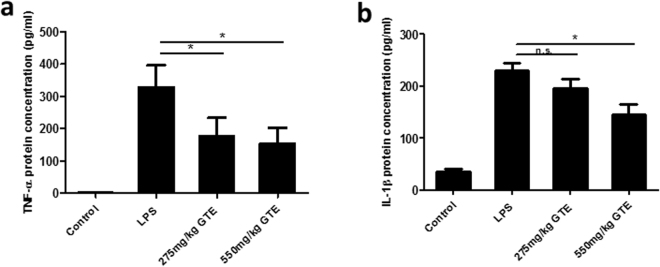
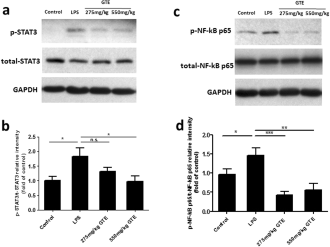
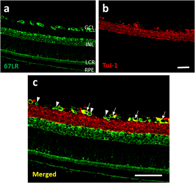
Inflammation is in a wide spectrum of retinal diseases, causing irreversible blindness and visual impairment. We have previously demonstrated that Green Tea Extract (GTE) is a potent anti-inflammatory agent for anterior uveitis. Here we investigated the anti-inflammatory effect of GTE on lipopolysaccharides (LPS)-induced retinal inflammation in rats and explored the underlying mechanism. Adult rats were injected with LPS and GTE was administered intra-gastrically at 2, 8, 26 and 32 hours post-injection. Staining of whole-mount retina showed that the number of activated microglia cells was significantly increased at 48 hours post-injection, which was suppressed after GTE treatment in a dose-dependent manner. Activation of astrocytes and Müller glia in the retina was also suppressed after GTE treatment. Meanwhile, GTE reduced the expression of pro-inflammatory cytokines including IL-1β, TNF-α and IL-6 in retina and vitreous humor. These anti-inflammatory effects were associated with a reduced phosphorylation of STAT3 and NF-κB in the retina. Furthermore, the surface receptor of EGCG, 67LR, was localized on the neurons and glia in the retina. These findings demonstrate that GTE is an effective agent in suppressing LPS-induced retinal inflammation, probably through its potent anti-oxidative property and a receptor-mediated action on transcription factors that regulate production of pro-inflammatory cytokines.


Conflict of interest statement

The authors declare that they have no competing interests.

Figures

Figure 1
Figure 1
Green tea extract suppressed the infiltrated cell number and protein accumulation in vitreous humor. (a,b) H&E sections showed that a large amount of infiltrated cells aggregated around the optic disc (OD) at 48 hrs after LPS stimuli. (c,d) Both 275 mg/kg GTE and 550 mg/ml GTE supplement after LPS stimulation decreased the cell number accumulated in vitreous humor (VH) dramatically. Scale bar 200 µm. (e) Analysis of vitreous humor demonstrated that the number of infiltrated cells and protein concentration was reduced dramatically in LPS injected rats after GTE administration. *p < 0.05, **p < 0.01, n = 7 in each group.
Figure 2
Figure 2
Green tea extract reduced the number of activated microglia in ganglion cell layer. (ac) Only a few OX-42 positive microglia cells and well-organized astrocytes network were observed in control retina. (df) After LPS treated, numerous microglia cells emerged in the retinal ganglion cell layer and the processes of astrocytes were disrupted. Meanwhile, some astrocytes became hypertrophied (arrowhead) and the endfeet of Müller glia cells (white arrow) appeared as well. Moreover, the glial endfeet along the capillary (C) were highly disorganized. GTE treatment at the dosage of 275 mg/kg (gi) and 550 mg/kg (jl) reduced the number of microglia cells and improved the disordered condition of astrocytes. The astrocytic array and blood-retinal barrier was preserved, especially under high dose group (k). Scale bar, 40 µm. (m) A total of 12 areas were imaged from each retina flat mount to calculate an average number of the immunoreactive microglia cells. (n) The number of activated microglia cells was significantly suppressed by the treatment of GTE in both 275 mg/kg and 550 mg/kg dosage. **p < 0.01, n = 6.
Figure 3
Figure 3
Green tea extract attenuated the activation of Müller glial cells. (ac) GFAP staining labelled a dense layer of astrocytes and a few Müller glia processes that run through the whole thickness of the retina in control group. (b) IBA-1 staining showed the localization of a few microglia in the ganglion cell layer (GCL) and inner nuclear layer (INL). (cf) After LPS insult, the expression of GFAP in Müller glia cells was elevated obviously, which accompanied by an increase in microglial cells in GCL and INL. (gl) GTE treatments attenuated the intensity of GFAP staining and density of IBA-1 staining dramatically, especially in high dose group. (m) Measurement of the fluorescence intensity in these tissues showed GTE produced a dose-dependent reduction of GFAP immunoreactivity. *p < 0.05, n = 6.
Figure 4
Figure 4
Green tea extract decreased the IL-1β, TNF-α, IL-6 and MMP9 gene expression in retina. (ac) The retina inflammatory-related gene expression was detected by quantitative-PCR. The gene expression of IL-1β, TNF-α and IL-6 were elevated by LPS and inhibited by GTE intake. (d) However, the anti-inflammatory gene, IL-10 did not show any significant change in both LPS and GTE group. (e) Moreover, MMP9, a gene that regulates the migration ability was down regulated in GTE group compared to LPS group. *p < 0.05, n.s. no significantly difference, n = 7.
Figure 5
Figure 5
Green tea extract inhibited the IL-1β, TNF-α protein accumulation in vitreous humor. (a,b) The concentrations of pro-inflammatory factors TNF-α, IL-1β were increased significantly 48 hrs after LPS treatment. 550 mg/kg GTE produced a prominent reduction on the secretion of these two cytokines compared LPS group. However, the lower dosage group (275 mg/kg group) only had a lower TNF-α concentration but no statistical differences from the LPS group in the concentration of IL-1β. *p < 0.05, n = 7.
Figure 6
Figure 6
Green tea extract inhibited the phosphorylation of STAT3 and NF- κB p65 in LPS-induced rat retina. (a,b) Western blot results showed that the phosphorylation of STAT3 and NF- κB p65 (Ser 536) were substantially increased in LPS treated rats. GTE treatment at 275 mg/kg and 550 mg/kg suppressed their phosphorylation level. (c,d) Quantitative measurements of the bands showed that treatment of GTE remarkably attenuated the phosphorylation of STAT3 and NF- κB p65. **p < 0.01, ***p < 0.001, n = 7.
Figure 7
Figure 7
The receptor of EGCG, 67LR, was expressed in the retina. (a) 67LR staining was observed in GCL, INL, layer of cones and rods (LCR) and retinal pigment epithelium layer (RPE). (b,c) Some of the 67LR staining in GCL was co-localized with TUJ1 staining (white arrow), which recognizes the neuronal class III β-Tubulin in retinal ganglion cells and their dendrites. Meanwhile, the 67LR staining was also observed on some relatively smaller cells (arrowhead) in GCL, which are likely the glial cells. Scale bars: 40 µm, n = 4.

References

    1. Pekal A, Drozdz P, Biesaga M, Pyrzynska K. Screening of the antioxidant properties and polyphenol composition of aromatised green tea infusions. J. Sci. Food Agric. 2012;92:2244–2249. doi: 10.1002/jsfa.5611. - DOI - PubMed
    1. Chan EW, Soh EY, Tie PP, Law YP. Antioxidant and antibacterial properties of green, black, and herbal teas of Camellia sinensis. Pharmacognosy. Res. 2011;3:266–272. doi: 10.4103/0974-8490.89748. - DOI - PMC - PubMed
    1. Seeram NP, et al. Catechin and caffeine content of green tea dietary supplements and correlation with antioxidant capacity. J. Agric. Food Chem. 2006;54:1599–1603. doi: 10.1021/jf052857r. - DOI - PubMed
    1. Cabrera C, Artacho R, Gimenez R. Beneficial effects of green tea–a review. J. Am. Coll. Cardiol. 2006;25:79–99. - PubMed
    1. Fujimura Y, et al. Green tea polyphenol EGCG sensing motif on the 67-kDa laminin receptor. PLoS One. 2012;7:e37942. doi: 10.1371/journal.pone.0037942. - DOI - PMC - PubMed

Publication types

MeSH terms

LinkOut - more resources