. 2018 Jan;25(1):101-108.
doi: 10.1038/s41594-017-0005-5. Epub 2017 Dec 11.

Histone octamer rearranges to adapt to DNA unwrapping

Affiliations

Histone octamer rearranges to adapt to DNA unwrapping

Silvija Bilokapic et al. Nat Struct Mol Biol. 2018 Jan.

Abstract

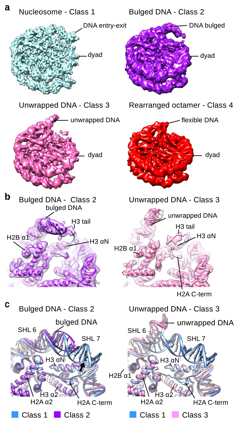
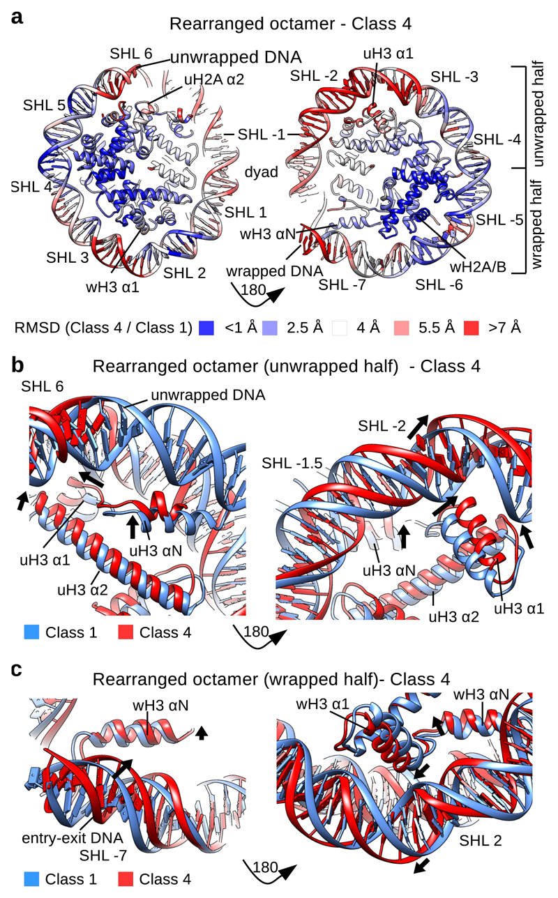
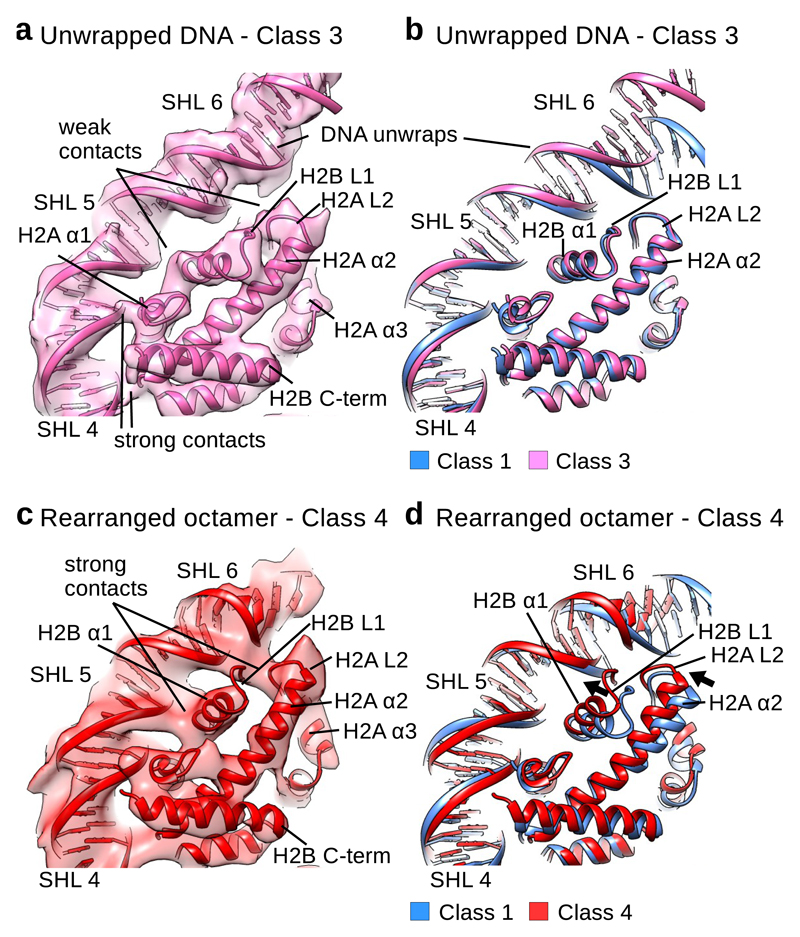
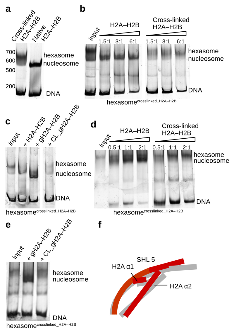
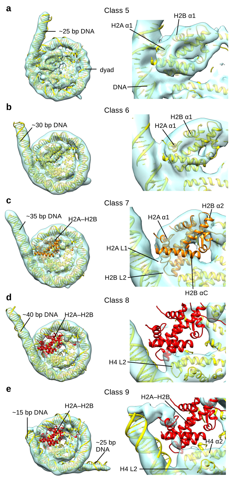
Nucleosomes, the basic units of chromatin, package and regulate expression of eukaryotic genomes. Although the structure of the intact nucleosome is well characterized, little is known about structures of partially unwrapped, transient intermediates. In this study, we present nine cryo-EM structures of distinct conformations of nucleosome and subnucleosome particles. These structures show that initial DNA breathing induces conformational changes in the histone octamer, particularly in histone H3, that propagate through the nucleosome and prevent symmetrical DNA opening. Rearrangements in the H2A-H2B dimer strengthen interaction with the unwrapping DNA and promote nucleosome stability. In agreement with this, cross-linked H2A-H2B that cannot accommodate unwrapping of the DNA is not stably maintained in the nucleosome. H2A-H2B release and DNA unwrapping occur simultaneously, indicating that DNA is essential in stabilizing the dimer in the nucleosome. Our structures reveal intrinsic nucleosomal plasticity that is required for nucleosome stability and might be exploited by extrinsic protein factors.


Conflict of interest statement

Competing financial interests. The authors declare no competing financial interests.

Figures

Fig. 1
Fig. 1. Cryo-EM reconstructions of NCP with unwrapped DNA.
a, Cryo-EM maps of the canonical NCP (Class 1) and NCPs with unwrapped DNA (Class 2-4). ~10 % of particles have unwrapped DNA at one entry-exit site. The Class 1 map (blue) was reconstructed to 3.7 Å (0.143 cutoff in FSC curve); the Class 2 map (purple) was reconstructed to 5.4 Å; the Class 3 map (pink) was reconstructed to 5.1 Å; the Class 4 map (red) was reconstructed to 6.3 Å. All maps are shown at similar contour levels. b, Fitting of the Class 2 (purple) and the Class 3 (pink) models into cryo EM maps. The X-ray structure of the NCP (PDB:3LZ1) was refined into the Class 2 and Class 3 cryo EM maps. In the Class 2 map the H3 tail binds the DNA and retains it at the histone octamer. In the Class 3 map, DNA is detached from the H3 tail. H2A C-terminal region is delocalized in the Class 3 structure. c, Comparison of the Class 1 model of NCP (blue) with Class 2 (purple) and Class 3 (pink) models. In the Class 2 structure DNA bulges but remains attached to the octamer. In the Class 3 structure entry-exit site DNA detaches from the octamer. This leads to delocalization of H2A C-terminal tail which binds the entry-exit site DNA. Other histones are in the canonical conformation.
Fig. 2
Fig. 2. Histone octamer rearranges when DNA unwraps.
a, RMSD (Cα) between the models for the Class 1 and the Class 4 structures showing the extent of rearrangements in the NCP. The DNA at SHL +- 2 and SHL +- 6-7 shows the largets movements between the Class 4 and canonical nucleosome structures. H3 αN, H3 α1 and α2, uH2A α2 and uH2B α1 show the largest rearrangements in the histone octamer. b, Conformational rearrangement of H3 in the half of the nucleosome with the unwrapped DNA. uH3 αN, uH3 α1 and uH3 α2 move in the Class 4 structure when compared to the Class 1 structure. DNA at SHL -2 moves outward. c, Conformational rearrangement of H3 in the half of the nucleosome with the wrapped DNA. wH3 αN and wH3 α1 move in the Class 4 structure when compared to the Class 1 structure. DNA at SHL 2 moves outward. wH3 αN and DNA at SHL -7 (second entry-exit site) move inward.
Fig. 3
Fig. 3. H2A–H2B dimer rearranges and binds unwrapping DNA.
a, Fitting of the Class 3 model into the cryo EM map. In the Class 3 cryo EM map H2B α1 (SHL 4.5), H2B L1 (H2B α1 and α2) and H2A L2 (H2A α2 and α3) (SHL 5.5) form weaker contacts with the DNA that are only visible at low contour level. H2A α1 at SHL 4.5 and H2A–H2B contacts at SHL 3.5 are similar to the contacts on the side with wrapped DNA. b, Comparison of the Class 1 model (blue) with the model for the Class 3 (pink) cryo EM map. In the Class 3 structure, H2A–H2B is in the conformation observed in the crystal structure. DNA at SHL 5 moves outward when compared to the Class 1 model. c, Fitting of the Class 4 model into the cryo EM map. In the Class 4 cryo EM map H2B α1 (SHL 4.5), H2B L1 (H2B α1 and α2) and H2A L2 (H2A α2 and α3) (SHL 5.5) form strong contacts with the DNA that are comparable to other contacts between histones and DNA. d, Comparison of the Class 1 model (blue) with the model for the Class 4 (red) cryo EM maps. In the Class 4 structure, H2B α1 and α2 and H2A α2 and α3 tilt toward the unwrapping DNA to stabilize the interaction.
Fig. 4
Fig. 4. Cross-linked H2A–H2B is not stably bound to the nucleosome.
a, Native gel showing nucleosome assembly with native and cross-linked H2A–H2B. When nucleosome were assembled with cross-linked H2A–H2B, primarily hexasomes were obtained. b, Native and cross-linked H2A–H2B were added to the assembled hexasomes (a) containing one cross-linked H2A–H2B. Native H2A–H2B is incorporated into hexasome forming the nucleosome. Cross-linked H2A–H2B is not stably incorporated into the hexasome. c, Native gel showing nucleosome assembly with native and cross-linked globular H2A–H2B. Native and cross-linked globular H2A–H2B were added to the previously assembled hexasomes containing one cross-linked H2A–H2B. Native full length and globular H2A–H2B are incorporated into hexasome forming the nucleosome. Cross-linked globular H2A–H2B is not stably incorporated into the hexasome. d, Native gel showing Nap1-mediated nucleosome assembly with native and cross-linked H2A–H2B. When we used cross-linked H2A–H2B, Nap1 also primarily assembled hexasomes (input). Native and cross-linked H2A–H2B were added to the assembled hexasomes. Nap1 incorporated native H2A–H2B into hexasomes forming nucleosomes. Nap1 was unable to incorporate cross-linked H2A–H2B into the hexasomes. e, Native gel showing Nap1-mediated nucleosome assembly with native and cross-linked globular H2A–H2B. Native and cross-linked globular H2A–H2B were added to the assembled hexasomes. Nap1 incorporated native globular H2A–H2B into hexasomes forming nucleosomes. Nap1 was unable to stably incorporate cross-linked globular H2A–H2B into the hexasomes. f, Unwrapping of DNA at SHL 5.5 induces conformational changes in H2A–H2B. Rearrangement of H2A α2 and α3 and H2B α1 maintains the interaction with the DNA and stabilizes the nucleosome. Representative images of at least 3 independent experiments are shown. Uncropped gel images are shown in Supplementary Data Set 1.
Fig. 5
Fig. 5. Cryo-EM reconstructions of distinct NCP and hexasome conformations.
a, Cryo-EM map of NCP (Class 5) with docked model (left). ~25 bp of DNA is not organized by the nucleosome in Class 5. A close view of the H2A–H2B dimer and the last contact with the DNA is shown on the right. b, Cryo-EM map of NCP (Class 6) with docked model (left). ~30 bp of DNA is not organized by the nucleosome in Class 6. A close view of the H2A–H2B dimer and the last contact with the DNA is shown on the right. c, Cryo-EM map of NCP (Class 7) with docked model (left). ~35 bp of DNA is not organized by the nucleosome in Class 7. A close view of the H2A–H2B dimer and last contact with the DNA is shown on the right. Only partial density could be observed for the H2A–H2B dimer, colored orange. d, Cryo-EM map of the hexasome (Class 8) with docked model (left). ~40 bp of DNA is not organized by the nucleosome in Class 8. A close view of the last contact with the DNA is shown on the right. No density for H2A–H2B dimer, colored red, could be observed in the map. e, Cryo-EM map of the hexasome (Class 9) with docked model (left). ~15 + ~25 bp of DNA is not organized by the nucleosome in Class 9. A close view of the last contact with the DNA is shown on the right. No density for the H2A–H2B dimer, colored red, could be observed in the map.

References

    1. Cutter AR, Hayes JJ. A brief review of nucleosome structure. FEBS Lett. 2015;589:2914–2922. - PMC - PubMed
    1. Andrews AJ, Luger K. Nucleosome structure(s) and stability: variations on a theme. Annu Rev Biophys. 2011;40:99–117. - PubMed
    1. Luger K, Mäder AW, Richmond RK, Sargent DF, Richmond TJ. Crystal structure of the nucleosome core particle at 2.8 A resolution. Nature. 1997;389:251–260. - PubMed
    1. Luger K, Dechassa ML, Tremethick DJ. New insights into nucleosome and chromatin structure: an ordered state or a disordered affair? Nat Rev Mol Cell Biol. 2012;13:436–447. - PMC - PubMed
    1. Li G, Levitus M, Bustamante C, Widom J. Rapid spontaneous accessibility of nucleosomal DNA. Nat Struct Mol Biol. 2005;12:46–53. - PubMed

Publication types

LinkOut - more resources