. 2018 Aug;39(8):1294-1304.
doi: 10.1038/aps.2017.143. Epub 2018 Jan 11.

Ulinastatin attenuates LPS-induced inflammation in mouse macrophage RAW264.7 cells by inhibiting the JNK/NF-κB signaling pathway and activating the PI3K/Akt/Nrf2 pathway

Affiliations

Ulinastatin attenuates LPS-induced inflammation in mouse macrophage RAW264.7 cells by inhibiting the JNK/NF-κB signaling pathway and activating the PI3K/Akt/Nrf2 pathway

Si-Tong Li et al. Acta Pharmacol Sin. 2018 Aug.

Abstract

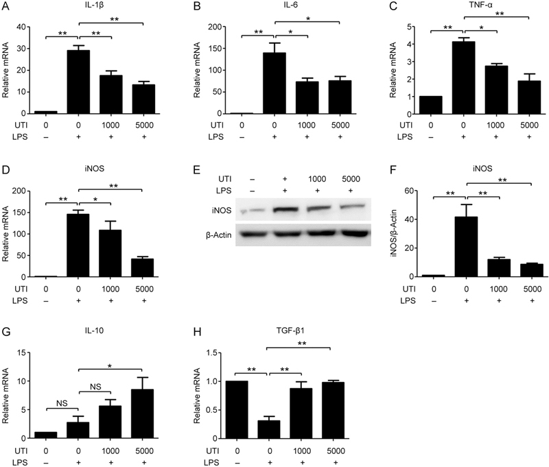
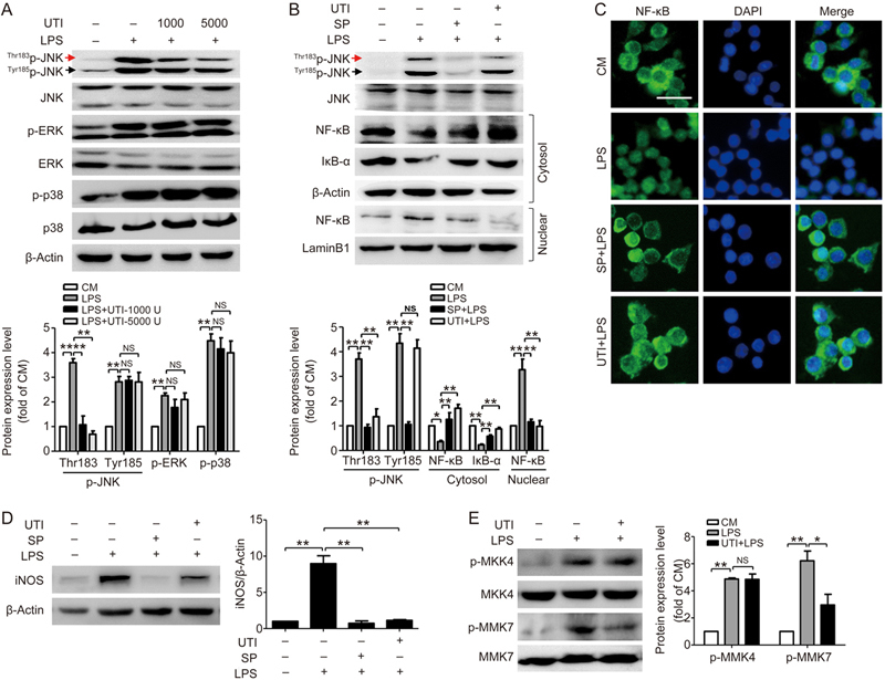
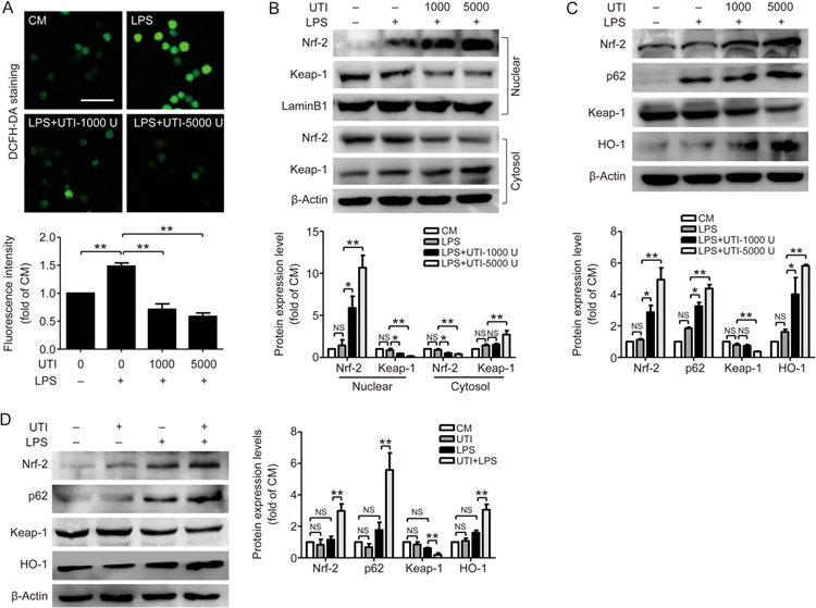
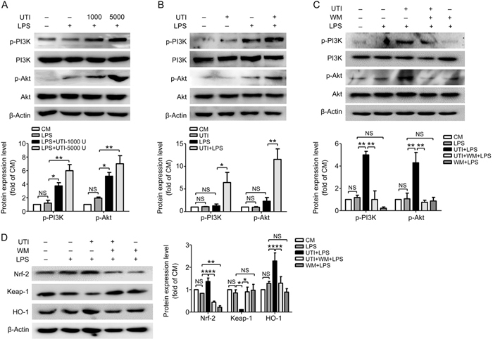
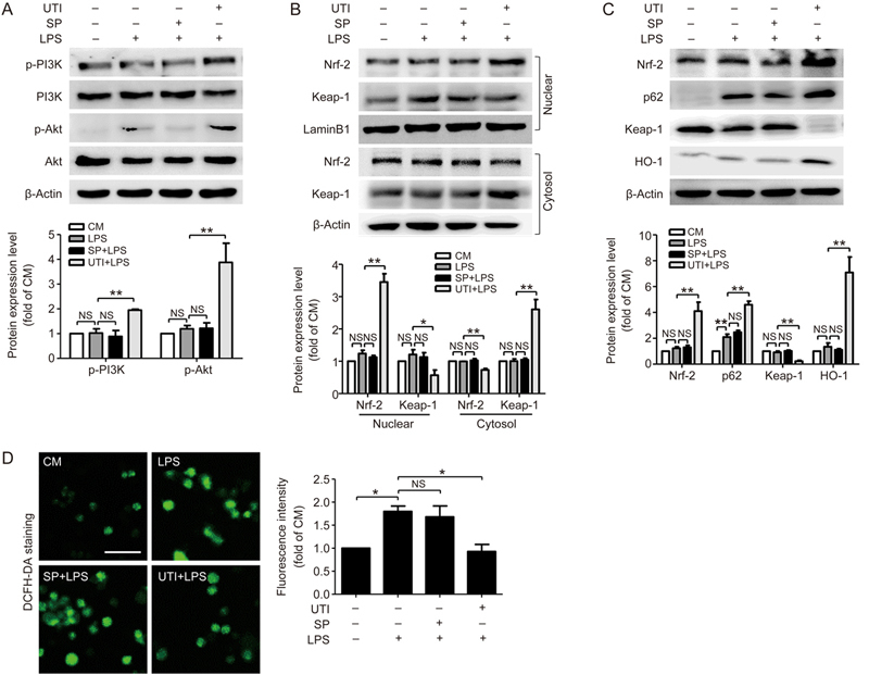
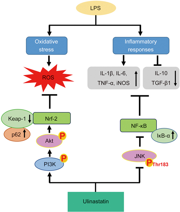
Ulinastatin (UTI) is a broad-spectrum serine protease inhibitor isolated and purified from human urine with strong anti-inflammatory and cytoprotective actions, which is widely used for the treatment of various diseases, such as pancreatitis and sepsis. Although the therapeutic effects of UTI are reported to be associated with a variety of mechanisms, the signaling pathways mediating the anti-inflammatory action of UTI remain to be elucidated. In the present study we carried out a systematic study on the anti-inflammatory and anti-oxidative mechanisms of UTI and their relationships in LPS-treated RAW264.7 cells. Pretreatment with UTI (1000 and 5000 U/mL) dose-dependently decreased the mRNA levels of pro-inflammatory cytokines (IL-1β, IL-6, TNF-α, iNOS) and upregulated anti-inflammatory cytokines (IL-10 and TGF-β1) in LPS-treated RAW264.7 cells. UTI pretreatment significantly inhibited the nuclear translocation of NF-κB by preventing the degradation of IκB-α. UTI pretreatment only markedly inhibited the phosphorylation of JNK at Thr183, but it did not affect the phosphorylation of JNK at Tyr185, ERK-1/2 and p38 MAPK; JNK was found to function upstream of the IκB-α/NF-κB signaling pathway. Furthermore, UTI pretreatment significantly suppressed LPS-induced ROS production by activating PI3K/Akt pathways and the nuclear translocation of Nrf2 via promotion of p62-associated Keap1 degradation. However, JNK was not involved in mediating the anti-oxidative stress effects of UTI. In summary, this study shows that UTI exerts both anti-inflammatory and anti-oxidative effects by targeting the JNK/NF-κB and PI3K/Akt/Nrf2 pathways.

Keywords: JNK; LPS; NF-κB; Nrf2; PI3K/Akt; RAW264.7 cells; ROS; cytokines; inflammation; ulinastatin.


Figures

Figure 1
Figure 1
UTI decreases pro-inflammatory cytokines and increases anti-inflammatory cytokines in LPS-treated RAW264.7 cells. Cells were pretreated with UTI (1000 or 5000 U/mL) for 3 h and then stimulated with LPS (1 μg/mL). After 6 h stimulation, the mRNA levels of IL-1β (A) and IL-6 (B) were measured by qRT-PCR. After 12 h stimulation, the mRNA levels of TNF-α (C), iNOS (D), IL-10 (G) and TGF-β1 (H) were analyzed by qRT-PCR, and the protein expression of iNOS (E–F) was detected by Western blot. The data are presented as the mean±SD of three independent experiments. * P<0.05, ** P<0.01. NS, not significant.
Figure 2
Figure 2
UTI suppresses the IκB-α/NF-κB signaling pathway in RAW264.7 cells. Cells were pretreated with or without UTI (1000, 5000 U/mL) for 3 h, followed by incubation with LPS (1 μg/mL) for 15 min (A, B) or 12 h (C). (A) Nuclear fractions and cytoplasmic fractions were extracted and subjected to immunoblot analysis using an anti-p65 antibody and an anti-IκB-α antibody. Lamin B1 and β-actin expression were measured to confirm equal protein amounts in nuclear extracts and cytoplasmic fractions. (B) The nuclear translocation of NF-κB p65 was visualized by immunofluorescence. The nuclei were counterstained with DAPI (blue). Scale bar=200 μm. (C) The total protein levels of NF-κB p65 and IκB-α were measured by Western blot. The data are presented as the mean±SD of three independent experiments. * P<0.05, ** P<0.01. NS, not significant.
Figure 3
Figure 3
UTI inhibits Thr183p-JNK. (A) The effects of UTI on the phosphorylation of JNK, ERK1/2 or p38 in LPS-treated RAW264.7 cells. Cells were incubated for 3 h with or without UTI (1000 or 5000 U/mL), then the cells were incubated with LPS (1 μg/mL) for 15 min, and aliquots of cell lysates containing equal amounts of protein were subjected to immunoblotting. (B–C) Cells were pretreated with or without UTI (5000 U/mL) for 3 h and SP600125 (SP, 10 μmol/L) for 1 h before co-incubation with LPS for 15 min. Nuclear fractions and cytoplasmic fractions were extracted and subjected to immunoblot analysis using an anti-p65 antibody and an anti-IκB-α antibody (B). The nuclear translocation of NF-κB p65 was observed by immunofluorescence. The nuclei were counterstained with DAPI (C). Scale bar = 200 μm. (D) After co-incubation with LPS for 12 h, the total protein levels of iNOS were measured by Western blot. (E) Cells were treated as (A). After co-incubation with LPS for 15 min, the total protein levels of p-MKK4 and p-MKK7 were measured by Western blot. The data are presented as the mean±SD of three independent experiments. * P<0.05, ** P<0.01. NS, not significant.
Figure 4
Figure 4
UTI protects against LPS-induced ROS stress. RAW264.7 cells were pretreated with or without UTI (1000 or 5000 U/mL) for 3 h, followed by exposure to LPS (1 μg/mL). (A) After 12 h, the products of ROS were detected using DCFH-DA, and the average fluorescence intensity from randomly selected fields (10 for each group) was measured. Scale bar=400 μm. (B) After 15 min, nuclear fractions and cytoplasmic fractions were extracted and subjected to Western blotting. (C) After 12 h, expression of Nrf2 and Keap1, as well as p62 and HO-1 was detected by Western blotting. (D) Cells were pre-incubated for 3 h with UTI (5000 U/mL) and were then treated with or without LPS for 12 h. The data are presented as the mean±SD of three independent experiments. * P<0.05, ** P<0.01. NS, not significant.
Figure 5
Figure 5
UTI activates the PI3K/Akt pathway in an LPS model. (A) RAW264.7 cells were pretreated with or without UTI (1000 or 5000 U/mL) for 3 h before exposure to LPS (1 μg/mL) for 15 min. Immunoblotting and densitometric analysis were performed. (B) Cells were pretreated with UTI (5000 U/mL) for 3 h, and then were stimulated with or without LPS for 15 min. (C) Cells were pretreated with or without UTI (5000 U/mL) for 3 h and wortmannin (10 μmol/L) for 1 h before LPS treatment for 15 min. Expression levels of p-PI3K, PI3K, p-Akt and Akt were detected by Western blot. (D) Cells were treated and (C), after treatment with LPS for 12 h, expression levels of Nrf2, Keap1 and HO-1 were detected. The data are presented as the mean ± SD of three independent experiments. *P<0.05; **P<0.01; NS, not significant.
Figure 6
Figure 6
The activation effect of UTI on the Nrf2 pathway is JNK-independent. (A–D) RAW264.7 cells were pretreated with or without UTI (5000 U/mL) for 3 h, and with or without SP600125 (10 μmol/L) for 1 h before co-incubating with LPS. After 15 min, (A) immunoblotting and densitometric analysis showed p-PI3K, PI3K, p-Akt and Akt protein levels in RAW264.7 cells. (B) Nuclear fractions and cytoplasmic fractions were extracted and subjected to Western blotting. (C) After 12 h, expression of Nrf2 and Keap1, as well as p62 and HO-1 was detected. (D) Cells were treated and (C), production of ROS was detected using DCFH-DA. Scale bar=400 μm. The data are presented as the mean±SD of three independent experiments. * P<0.05; ** P<0.01. NS, not significant.
Figure 7
Figure 7
Scheme summarizing the protective effects of UTI on LPS-induced RAW264.7 cells. Ulinastatin (UTI) treatment effectively suppressed LPS-induced inflammatory injury. This is largely dependent on the suppression of LPS-activated JNK phosphorylation at Thr183, thereby inhibiting IκBα down-regulation and the NF-κB signaling pathway. In addition, UTI performed its role in anti-inflammation through ROS suppression, which mainly relies on the upregulation of the Nrf2-mediated antioxidation pathway by promoting p62-associated Keap1 degradation and the PI3K/Akt pathway. Meanwhile, the JNK signaling pathway is not involved in mediating the Nrf2 pathway.

References

    1. Inoue K, Takano H. Urinary trypsin inhibitor as a therapeutic option for endotoxin-related inflammatory disorders. Expert Opin Invest Drugs. 2010;19:513–20. doi: 10.1517/13543781003649533. - DOI - PubMed
    1. Linder A, Russell JA. An exciting candidate therapy for sepsis: ulinastatin, a urinary protease inhibitor. Intensive Care Med. 2014;40:1164–7. doi: 10.1007/s00134-014-3366-9. - DOI - PubMed
    1. Pan Y, Fang H, Lu F, Pan M, Chen F, Xiong P, et al. Ulinastatin ameliorates tissue damage of severe acute pancreatitis through modulating regulatory T cells. J Inflammation. 2017;14:7. doi: 10.1186/s12950-017-0154-7. - DOI - PMC - PubMed
    1. Zhang H, Tan C, Wang X, Kang D, Chen Y, Xiong J, et al. Preventive effects of ulinastatin on complications related to pancreaticoduodenectomy: a Consort-prospective, randomized, double-blind, placebo-controlled trial. Medicine. 2016;95:e3731. doi: 10.1097/MD.0000000000003731. - DOI - PMC - PubMed
    1. Song D, Song G, Niu Y, Song W, Wang J, Yu L, et al. Ulinastatin activates haem oxygenase 1 antioxidant pathway and attenuates allergic inflammation. Br J Pharmacol. 2014;171:4399–412. doi: 10.1111/bph.12780. - DOI - PMC - PubMed

MeSH terms