Review
. 2018 Mar;14(3):146-156.
doi: 10.1038/nrrheum.2017.213. Epub 2018 Jan 11.

Updating osteoimmunology: regulation of bone cells by innate and adaptive immunity

Affiliations
Review

Updating osteoimmunology: regulation of bone cells by innate and adaptive immunity

Matthew C Walsh et al. Nat Rev Rheumatol. 2018 Mar.

Abstract

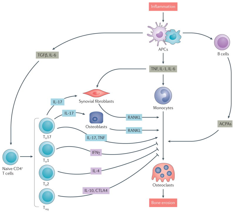
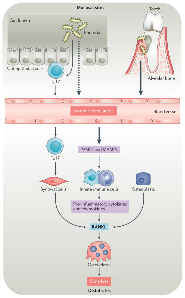
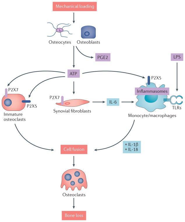
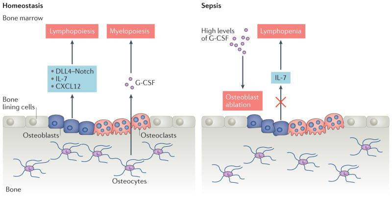
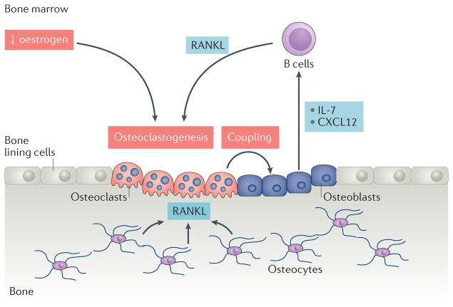
Osteoimmunology encompasses all aspects of the cross-regulation of bone and the immune system, including various cell types, signalling pathways, cytokines and chemokines, under both homeostatic and pathogenic conditions. A number of key areas are of increasing interest and relevance to osteoimmunology researchers. Although rheumatoid arthritis has long been recognized as one of the most common autoimmune diseases to affect bone integrity, researchers have focused increased attention on understanding how molecular triggers and innate signalling pathways (such as Toll-like receptors and purinergic signalling pathways) related to pathogenic and/or commensal microbiota are relevant to bone biology and rheumatic diseases. Additionally, although most discussions relating to osteoimmune regulation of homeostasis and disease have focused on the effects of adaptive immune responses on bone, evidence exists of the regulation of immune cells by bone cells, a concept that is consistent with the established role of the bone marrow in the development and homeostasis of the immune system. The active regulation of immune cells by bone cells is an interesting emerging component of investigations that seek to understand how to control immune-associated diseases of the bone and joints.


Conflict of interest statement

Competing interests statement

The authors declare no competing interests.

Figures

Figure 1
Figure 1. Immune regulation of bone destruction in rheumatoid arthritis (RA)
The interaction of immune cells with bone cells in RA leads to destructive inflammation that involves the activation of cytokine-mediated pathways that result in bone remodelling. Autoimmune responses are induced by inflammation: T cells and B cells are activated by antigen presenting cells (APCs) such as dendritic cells and macrophages in the inflamed synovium. Synovial fibroblasts produce the osteoclastogenic cytokine receptor activator of nuclear factor-κB ligand (RANKL), and CD4+ T cells produce osteoclastogenic cytokines such as IL 17 and TNF (produced by T helper 17 (TH17) cells) but also produce anti osteoclastogenic cytokines such as IFNγ (TH1 cells), IL 4 (TH2 cells) and IL 10 and cytotoxic T lymphocyte protein 4 (CTLA4; produced by regulatory T (Treg) cells). TH17 cells also stimulate osteoblastogenic activity by producing IL 17. Anti citrullinated protein antibodies (ACPAs) are produced by B cells in the RA joint and are also able to promote osteoclastogenesis and osteoclast activity. TGFβ, transforming growth factor β.
Figure 2
Figure 2. Microbiota and osteoimmunology
Under homeostatic or inflammatory conditions, microbiota can affect bone resorption at sites proximal to and distal to the site of colonization. Inflammatory conditions are often triggered by dysbiosis of microbiota, which can be associated with the outgrowth of particular phyla and species. Molecular triggers from gut microbiota cross the epithelial boundary more readily under dysbiotic and/or inflammatory conditions than during homeostasis and might eventually influence bone at distal sites, either by entering systemic circulation or by activating local adaptive immune elements, such as T helper 17 (TH17) cells within the mucosal tissue that can transit to distal sites and therein exert their effector function. By contrast, stimuli from microbiota in the subgingival crest between the tooth and the gum act proximally on the alveolar bone that anchors teeth. Oral microbiota can also contribute to inflammatory bone loss at distal sites, such as the joints of patients with rheumatoid arthritis. Pathogen-associated molecular patterns (PAMPs) and microbiota-associated molecular patterns (MAMPs) trigger signalling pathways in innate immune cells and synovial fibroblasts that induce the production of pro-inflammatory cytokines and chemokines. These factors contribute to increased expression of receptor activator of nuclear factor-κB ligand (RANKL) and increased osteoclast differentiation and activation, which increase inflammatory bone loss at both distal sites and proximal sites.
Figure 3
Figure 3. Purinergic signalling regulates bone cells and inflammation
ATP signals through members of the P2X receptor family of ligand-gated ion channels. Mechanical loading regulates the release of ATP and prostaglandin E2 (PGE2) by osteoblasts and osteocytes. Extracellular ATP signals through P2X7 or P2X5 on immature osteoclasts, triggering cell fusion and resulting in multi-nucleated osteoclasts. ATP also triggers the production of pro inflammatory cytokines such as IL 6 by synovial fibroblasts, and can synergize with lipopolysaccharide (LPS) signalling through Toll like receptors (TLRs) to activate inflammasomes and produce mature forms of IL 1β and IL 18 that also promote osteoclast multi nucleation and maturation. In an experimental setting, LPS triggered inflammatory bone loss requires purinergic signalling through the P2X5 receptor.
Figure 4
Figure 4. Interaction between immune cells and bone cells in homeostasis and sepsis
Under homeostatic conditions, osteolineage cells (osteoblasts and osteocytes) provide niches for haematopoiesis, lymphopoiesis and myelopoiesis to occur by producing cytokines and chemokines. Osteoclasts contribute to the provision of a favourable niche by maintaining the bone marrow microenvironment. By contrast, sepsis induces aberrant production of granulocyte colony stimulating factor (G CSF), which suppresses mature osteoblasts, resulting in the depletion of IL 7, an important lymphopoietic factor, and leading to lymphopenia. CXCL12, CXC motif chemokine 12; DLL4, Delta like protein 4.
Figure 5
Figure 5. The effects of tissue-specific RANKL on bone and the immune system
Sex hormone deficiency induces aberrant osteoclastogenesis owing to a lack | of inhibition provided by oestrogen. Increased levels of osteoclastogenesis lead to enhanced osteoblastic activity via coupling, a homeostatic process in which bone resorption and new bone formation are temporally and spatially coordinated, which results in the production of chemokines and cytokines that increase the generation of B cells. B cell derived receptor activator of nuclear factor κB ligand (RANKL) can further enhance osteoclastogenesis. CXCL12, CXC motif chemokine 12.

References

    1. Arron JR, Choi Y. Bone versus immune system. Nature. 2000;408:535–536. - PubMed
    1. Walsh MC, et al. Osteoimmunology: interplay between the immune system and bone metabolism. Annu Rev Immunol. 2006;24:33–63. - PubMed
    1. Yu VW, Scadden DT. Hematopoietic stem cell and its bone marrow niche. Curr Top Dev Biol. 2016;118:21–44. - PMC - PubMed
    1. Ash P, Loutit JF, Townsend KM. Osteoclasts derived from haematopoietic stem cells. Nature. 1980;283:669–670. - PubMed
    1. Walsh MC, Choi Y. Biology of the RANKL-RANK-OPG system in immunity, bone, and beyond. Front Immunol. 2014;5:511. - PMC - PubMed

Publication types

LinkOut - more resources