Review
. 2018 Jan 10;23(1):14-26.
doi: 10.1016/j.chom.2017.12.004.

Getting the "Kill" into "Shock and Kill": Strategies to Eliminate Latent HIV

Affiliations
Review

Getting the "Kill" into "Shock and Kill": Strategies to Eliminate Latent HIV

Youry Kim et al. Cell Host Microbe. .

Abstract

Despite the success of antiretroviral therapy (ART), there is currently no HIV cure and treatment is life long. HIV persists during ART due to long-lived and proliferating latently infected CD4+ T cells. One strategy to eliminate latency is to activate virus production using latency reversing agents (LRAs) with the goal of triggering cell death through virus-induced cytolysis or immune-mediated clearance. However, multiple studies have demonstrated that activation of viral transcription alone is insufficient to induce cell death and some LRAs may counteract cell death by promoting cell survival. Here, we review new approaches to induce death of latently infected cells through apoptosis and inhibition of pathways critical for cell survival, which are often hijacked by HIV proteins. Given advances in the commercial development of compounds that induce apoptosis in cancer chemotherapy, these agents could move rapidly into clinical trials, either alone or in combination with LRAs, to eliminate latent HIV infection.

Keywords: Akt inhibitors; Bcl-2 antagonists; HIV cure; HIV latency; PI3K inhibitors; RIG-I inducers; Smac mimetics; XIAP inhibitors; apoptosis; latency reversing agents; pro-apoptotic drugs; shock and kill.


Conflict of interest statement

Declaration of Interests

SL’s institution has received funding for investigator-initiated industry-sponsored studies from Merck, Gilead, Viiv Healthcare and Tetralogic, for educational activities from Merck, Viiv and Gilead, and has also acted on the advisory board for and as consultancy to Callimune, Tetralogic, and InnaVirVax. SL, JA and YK collaborate with Infinity Pharmaceuticals to test the impact of compounds on HIV latency. There are no other conflicts of interest.

Figures

Figure 1
Figure 1. Shock and Kill strategy to eliminate HIV latently infected cells
The “shock and kill” strategy uses latency reversing agents (LRAs) to increase HIV transcription, protein expression and virion production. The cell may potentially die through virus-mediated cytopathic events or immune-mediated clearance. LRAs: Latency reversing agents, ART: antiretroviral therapy
Figure 2
Figure 2. Classes of Latency reversing agents (LRAs)
Latency reversing agents (LRAs) can act on different pathways resulting in an increase in HIV transcription and/or virion production. P-TEFb: Positive transcription elongation factor b, TLR: Toll-like Receptor, mTOR: Mechanistic target of rapamycin, STAT5: Signal transducer and activator of transcription 5, IL-15: Interleukin-15
Figure 3
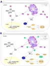
Figure 3. Effects of HIV proteins on apoptosis and compounds to foster apoptosis of cells
When stress stimuli induce the intrinsic mitochondrial pathway, the Bak or Bax members of the B-cell lymphoma 2 (Bcl-2) family are activated to promote apoptosis. However, activation of the PI3K pathway promotes cell survival by preventing steps in the apoptosis pathway. PI3K activation leads to conversion of phosphatidylinositol 4,5-biphosphate (PIP2) to phosphatidylinositol (3,4,5)-triphosphate (PIP3). This step is also negatively regulated by phosphatase and tensin homolog (PTEN). The conversion of PIP2 to PIP3 leads to the activation of Akt protein. Akt can inhibit the forkhead box protein O1 (FOXO1) transcription factor from translocating into the nucleus to induce pro-apoptotic target genes, preventing apoptosis. Additionally, Akt inhibits the pro-apoptotic protein Bad from inhibiting anti-apoptotic Bcl-2 members like Bcl-2 and Bcl-XL, which in turn is inhibited from activating Bak or Bax thereby also preventing apoptosis. A) Pathways leading to cell survival. Anti-apoptotic HIV proteins (shown in red) expressed early in the virus life cycle that can promote cell survival include Nef, Tat and Vpr. Nef can bind PI3K proteins to increase the conversion of PIP2 to PIP3. This leads to an increase in Akt protein that subsequently leads to inhibition of pro-apoptotic molecules such as Bad from activating apoptosis thereby promoting cell survival. Additionally, Nef can prevent p53 from activating the pro-apoptotic molecule PUMA which directly inhibits Bcl-2 to prevent apoptosis. HIV Tat protein can inhibit PTEN, thereby promoting the PI3K/Akt pathway to promote cell survival. Tat can also directly inhibit FOXO1 transcription factors to prevent the transcription of pro-apoptotic genes, preventing apoptosis. Tat also prevents the pro-apoptotic molecule PUMA from inhibiting Bcl-2. HIV Vpr can lead to the increase of anti-apoptotic molecule Bcl-2 in addition to inhibiting the pro-apoptotic Bak molecule. B) Pathways leading to cell death. When activated, Bax proteins form pores in the mitochondrial membrane, leading to the release of cytochrome C and second mitochondria-derived activator of caspase (Smac) into the cytosol. Cytosolic cytochrome C leads to the formation of the apoptosome that activates caspase-9. Caspase-9 then cleaves pro-caspase -3, -7 into their active caspase-3 and caspase-7 forms, which lead to apoptosis. Multiple HIV proteins (shown in green) can interact with the members of the apoptosis pathway later in the viral life cycle, leading to increased apoptosis. Nef can inhibit both the anti-apoptotic molecules Bcl-2 and Bcl-XL. Higher expression of HIV Vpr can bind and lead to the upregulation of Bak to initiate apoptosis. Higher expression of Tat also leads to the upregulation of Bax, resulting in the release of cytochrome C from the mitochondria and subsequent activation of the caspase cascade. HIV Env protein can inhibit the anti-apoptotic protein Bcl-2 to favour apoptosis. The HIV protease protein also cleaves caspase-8 to the pro-apoptotic Casp8p41 peptide to promote apoptosis. It is likely that the balance of pro-apoptotic versus anti-apoptotic cellular and viral proteins decide the fate of infected cells. C) Modulation of apoptosis pathways using compounds. Compounds (in light blue) that act on different cellular proteins in these pathways could potentially be used to tip the balance toward apoptosis of HIV infected cells. PI3K inhibitors block the conversion of PIP2 to PIP3, decreasing active Akt within a cell to stop Akt from inhibiting apoptosis. Akt inhibitors directly decrease active Akt to prevent Akt from inhibiting apoptosis to ultimately induce apoptosis. Bcl-2 inhibitors such as Venetoclax inhibit anti-apoptotic Bcl-2 to sensitize the cells towards apoptosis. Smac mimetics competitively bind inhibitors of apoptosis proteins like X-linked inhibitor of apoptosis (XIAP) to promote apoptosis. Retinoic acid-inducible gene I (RIG-1) detects viral RNA as well as activating pro-apoptotic Bak/Bax proteins, leading to apoptosis. In addition to viral RNA, RIG-1 inducer compounds like the retinoic acid derivative acitretin can also trigger apoptosis.

References

    1. Abbas W, Herbein G. Molecular Understanding of HIV-1 Latency. Adv Virol. 2012;2012:574967. - PMC - PubMed
    1. Adams JM, Cory S. The Bcl-2 protein family: arbiters of cell survival. Science. 1998;281(5381):1322–6. - PubMed
    1. Aillet F, Masutani H, Elbim C, Raoul H, Chene L, Nugeyre MT, Paya C, Barre-Sinoussi F, Gougerot-Pocidalo MA, Israel N. Human immunodeficiency virus induces a dual regulation of Bcl-2, resulting in persistent infection of CD4(+) T- or monocytic cell lines. J Virol. 1998;72(12):9698–705. - PMC - PubMed
    1. Amaravadi RK, Schilder RJ, Martin LP, Levin M, Graham MA, Weng DE, Adjei AA. A Phase I Study of the SMAC-Mimetic Birinapant in Adults with Refractory Solid Tumors or Lymphoma. Mol Cancer Ther. 2015;14(11):2569–75. - PubMed
    1. Annis MG, Soucie EL, Dlugosz PJ, Cruz-Aguado JA, Penn LZ, Leber B, Andrews DW. Bax forms multispanning monomers that oligomerize to permeabilize membranes during apoptosis. EMBO J. 2005;24(12):2096–103. - PMC - PubMed

MeSH terms