. 2018 Apr 2;128(4):1384-1396.
doi: 10.1172/JCI97611. Epub 2018 Feb 26.

Epithelial-mesenchymal transition leads to NK cell-mediated metastasis-specific immunosurveillance in lung cancer

Affiliations

Epithelial-mesenchymal transition leads to NK cell-mediated metastasis-specific immunosurveillance in lung cancer

Peter J Chockley et al. J Clin Invest. .

Abstract

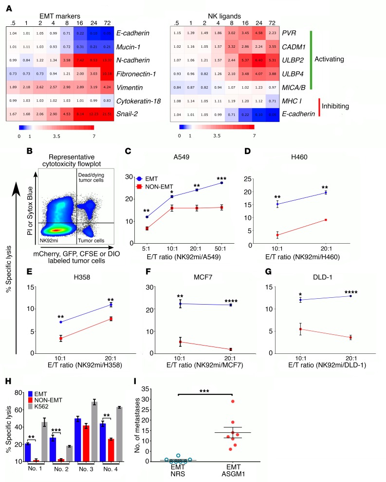
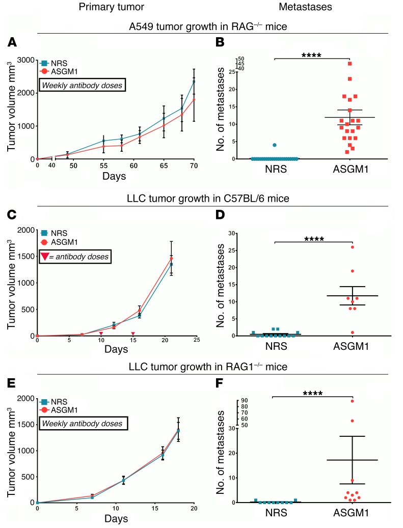
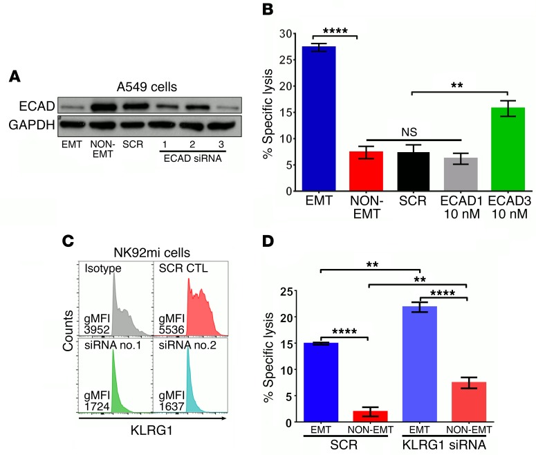
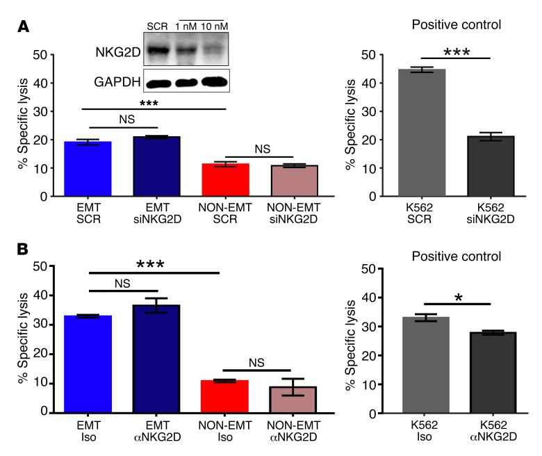
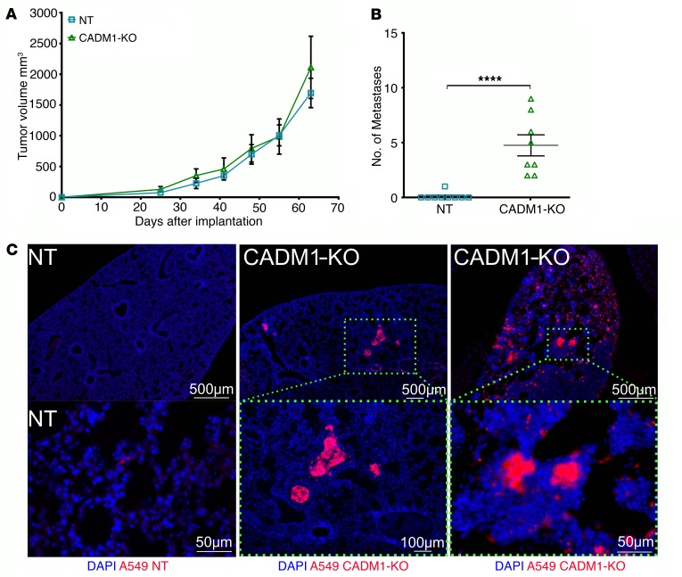
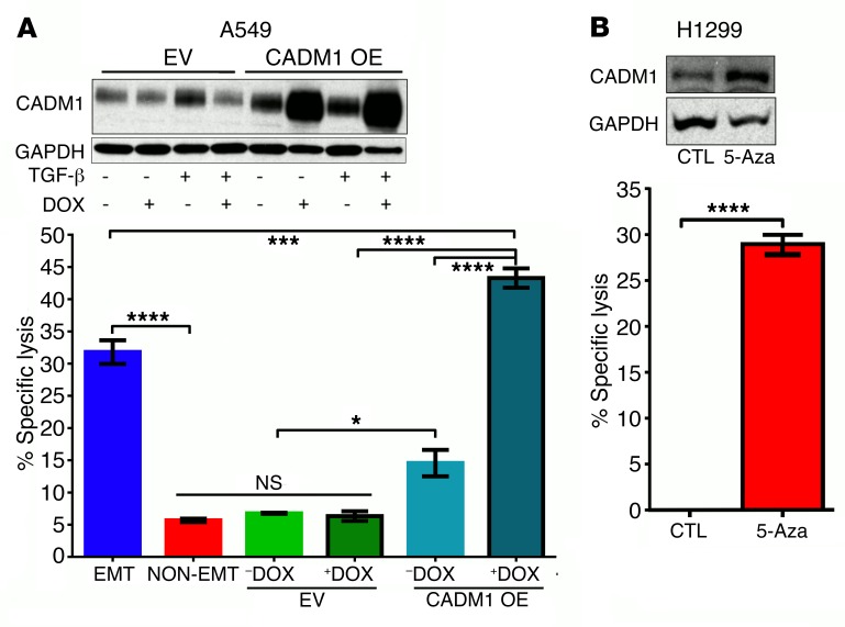
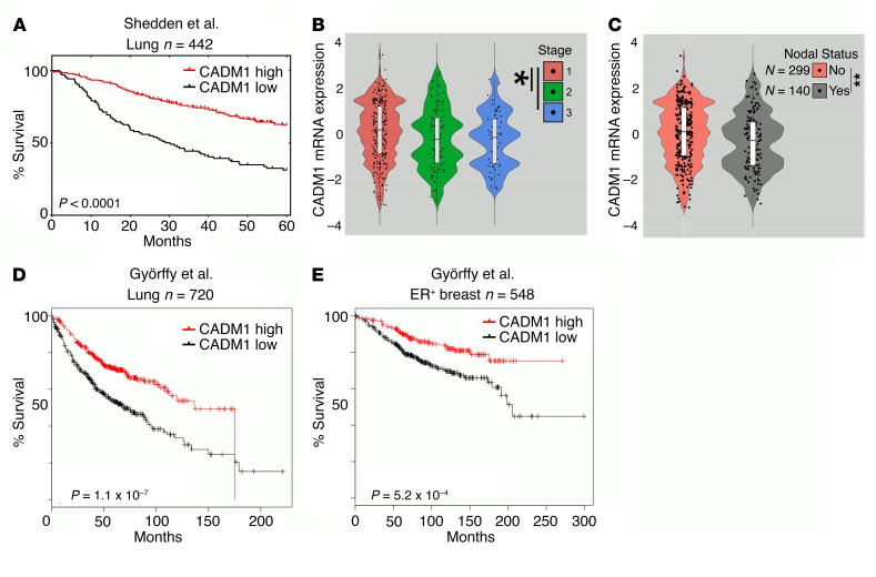
During epithelial-mesenchymal transition (EMT) epithelial cancer cells transdifferentiate into highly motile, invasive, mesenchymal-like cells, giving rise to disseminating tumor cells. Few of these disseminated cells successfully metastasize. Immune cells and inflammation in the tumor microenvironment were shown to drive EMT, but few studies investigated the consequences of EMT for tumor immunosurveillance. In addition to initiating metastasis, we demonstrate that EMT confers increased susceptibility to natural killer (NK) cells and contributes, in part, to the inefficiency of the metastatic process. Depletion of NK cells allowed spontaneous metastasis without affecting primary tumor growth. EMT-induced modulation of E-cadherin and cell adhesion molecule 1 (CADM1) mediated increased susceptibility to NK cytotoxicity. Higher CADM1 expression correlates with improved patient survival in 2 lung and 1 breast adenocarcinoma patient cohorts and decreased metastasis. Our observations reveal a novel NK-mediated, metastasis-specific immunosurveillance in lung cancer and present a window of opportunity for preventing metastasis by boosting NK cell activity.

Keywords: Immunology; Immunotherapy; Lung cancer; NK cells; Oncology.


Conflict of interest statement

Conflict of interest: The authors have declared that no conflict of interest exists.

Figures

Figure 1
Figure 1. EMT differentially regulates NK ligands and promotes susceptibility to NK-mediated cytotoxicity.
(A) Heatmap (blue: downregulation; red: upregulation) representing fold changes, from 0 hours to 72 hours, time course of differentially expressed EMT markers and NK ligand genes during TGF-β–induced EMT, from a previously published gene expression profile data set (GEO GSE17708) (36). (B) Representative flow cytometric plot of cytotoxicity assay showing locations of effector, NK92mi (fluorophore-null cells), and target cells (fluorophore positive) and their exclusion DNA-binding dye status (viability indicator). PI, propidium iodide. (CG) NK92mi-mediated cytotoxicity plots after 4 hours of coculture at indicated E/T ratios per cell type and treatment. Cell lines were treated with TGF-β (5 ng/ml) for 3, 6, 12, 6, and 6 days to induce optimum EMT, as assessed by complete E-cad downregulation and induction of vimentin or N-cadherin, in A549 (C), H460 (D), H358 (E), MCF7 (F), and DLD-1 (G), respectively. Data represent triplicate mean ± SEM, and 2-tailed unpaired t tests were performed. All experiments were repeated at least twice. (H) Freshly isolated human peripheral blood–derived NK cells were used as effector cells (E/T, 10:1) against A549 cells. K562 cells were used as a positive control for cytotoxicity. Data represent mean ± SEM, and 2-tailed, unpaired, t tests were performed. (I) To assess experimental metastasis, A549 cells were treated with TGF-β (5 ng/ml) in vitro and injected through the tail vein into RAG–/– mice. After 8 weeks, lungs were harvested to assess tumor burden. NRS, normal rabbit serum. Data represent 2 independent experiments, n = 3–4 for each group, and pooled results are shown. Error bars are SEM; Mann-Whitney U test was performed, *P < 0.05, **P < 0.01, ***P < 0.001, ****P < 0.0001.
Figure 2
Figure 2. NK cell depletion allows spontaneous metastatic spread without affecting primary tumor growth.
(AF) To assess the effect of NK cell depletion on primary tumor growth and metastasis, indicated cell lines were implanted subcutaneously under the dorsal flanks of RAG1–/– or C57BL/6 mice. Mice were treated weekly with anti–asialo GM1 antibody (ASGM1) to deplete NK cells or with normal rabbit serum (NRS) as control. (A, C, and E) Primary tumor growth was monitored, and mean tumor volumes are plotted with error bars as SEM. Representative data from a single experiment of at least duplicates. (B, D, and F) Overt lung nodules were counted on the excised lungs to assess spontaneous metastasis. Mouse strains and tumor cell implants are designated. Error bars are SEM; Mann-Whitney U test was performed, *P < 0.05, **P < 0.01, ***P < 0.001, ****P < 0.0001. Data represent at least 2 experiments, n = 4–5 per group, and pooled data are shown.
Figure 3
Figure 3. Loss of E-cad expression sensitizes tumor cells to NK-mediated cytotoxicity through KLRG1.
(A) A549 cells were transfected with 10 nM of scrambled (SCR) or 3 different E-cad–specific siRNA molecules. After 24 hours, cells were treated with (EMT) or without (NON-EMT) TGF-β (5 ng/ml) for 72 hours. E-cad and GAPDH expressions were assessed by Western immunoblotting. (B) Susceptibility to NK cytotoxicity was assessed using NK92mi cells as effectors, as described for Figure 1. Mean ± SEM is shown; 1-way ANOVA with Tukey’s post hoc analysis was performed, **P < 0.01, ****P < 0.0001. (C) NK92mi cells were transfected with 10 nM of SCR or KLRG1-specific siRNA. After 72 hours, KLRG1 expression was assessed by flow cytometry. gMFI, geometric mean fluorescence intensity. (D) NK92mi cells from C were used as effectors against EMT or non-EMT A549 cells in the NK cytotoxicity assay. Mean ± SEM is shown; 2-way ANOVA with Tukey’s post hoc analysis was performed, **P < 0.01, ****P < 0.0001. All experiments were repeated twice, and data are representative of 1 experiment.
Figure 4
Figure 4. NKG2D receptor is not involved in EMT-induced susceptibility to NK-mediated cytotoxicity.
(A) NK92mi cells were transfected with 10 nM of scrambled (SCR) or NKG2D-specific siRNA. After 72 hours, NKG2D expression was assessed by Western immunoblotting (inset), and these cells were used as effectors against A549 cells that were treated with (EMT) or without (NON-EMT) TGF-β (5 ng/ml) for 72 hours in the NK cytotoxicity assay. (B) NKG2D receptors were neutralized by treatment of NK92mi cells with anti-NKG2D receptor antibody (50 μg/ml) 45 minutes before coculturing with EMT or non-EMT A549 cells in the NK cytotoxicity assay. Iso, isotype control. All experiments were repeated twice; representative data of 1 experiment are shown. Mean ± SEM is shown; 2-way ANOVA with Tukey’s post hoc analysis was performed, *P < 0.05, ***P < 0.001. For positive control K562 cytotoxicity, mean ± SEM is shown; 2-tailed, unpaired t tests were performed, *P < 0.05, ***P < 0.001.
Figure 5
Figure 5. CADM1 expression is modulated by EMT-MET cycling and mediates tumor cell susceptibility to NK cytotoxicity.
(A) A549 cells were treated with TGF-β (5 ng/ml) to induce EMT, and total proteins were extracted at the indicated times. After 72 hours, cells were washed 3 times and replaced with fresh media to induce mesenchymal-epithelial transition (MET), and total proteins were extracted at the indicated times. Protein expression of E-cad, CADM1, vimentin, and GAPDH was assessed by Western immunoblotting. (B) A549 cells treated with and without TGF-β for 72 hours were fixed and assessed for E-cad and CADM1 expression by immunofluorescence staining. Scale bars: 100 μm. (C) To stably knock out CADM1, Cas9-expressing A549 cells were transduced with lentiviruses expressing 3 different CADM1-specific CRISPR sgRNAs and a nontargeting (NT) control sgRNA. CADM1 knockout was assessed by Western immunoblotting using 2 different CADM1 antibodies raised against C-terminal (CADM1-c) and N-terminal (CADM1-n) portions. (D and E) Susceptibility of CADM1-KO A549 cells to NK cytotoxicity was assessed by coculturing with either NK92mi cells or primary human blood–derived NK cells from 4 different donors. In D, mean ± SEM is shown; 2-way ANOVA with Tukey’s post hoc analysis was performed, **P < 0.01, ****P < 0.0001. In E, EMT controls are from Figure 1H, as these experiments were performed simultaneously. Mean ± SEM is shown; 2-tailed, unpaired t tests were performed, *P < 0.05, **P < 0.01.
Figure 6
Figure 6. Inhibition of CADM1 in tumor cells allows spontaneous metastasis without affecting primary tumor growth.
(A) mCherry-expressing CADM1 KO and control A549 cells were subcutaneously implanted into the dorsal flanks of RAG1–/– mice. Primary tumor growth was monitored, and mean tumor volumes are plotted; mean ± SEM shown. Data are representative of 1 experiment (n = 4–5 mice per group). (B) Overt lung nodules were counted on the excised lungs to assess spontaneous metastasis. Data represent 2 independent experiments, and pooled data are shown; error bars are SEM, and Mann-Whitney U test was performed, ****P < 0.0001. (C) Presence, or lack thereof, of metastatic spread was further confirmed by visualization of mCherry-positive tumor cells in the cross sections of the lungs by immunofluorescence. Top row scale bars: 500 μm; lower row: 50 μm, 100 μm, and 50μm, respectively.
Figure 7
Figure 7. Restoring CADM1 expression in tumor cells is sufficient to confer susceptibility to NK cytotoxicity.
(A) Stable A549 cell lines expressing empty vector (EV) or vector with doxycycline-inducible (DOX-inducible) human CADM1 overexpression (CADM1 OE) were developed. Expression of TGF-β–induced CADM1 was assessed in the presence and absence of doxycycline by Western immunoblotting, and susceptibility to NK cytotoxicity was assessed using NK92mi cells as effectors, as described for Figure 1. Mean ± SEM is shown; 1-way ANOVA with Tukey’s post hoc analysis was performed, *P < 0.05, ***P < 0.001, ****P < 0.0001. (B) H1299 cells harboring CADM1 promoter hypermethylation were cultured in the presence and absence of a pan–DNA methylase inhibitor, 5-azadeoxycytidine (5-Aza). CADM1 expression and NK cytotoxicity were assessed as described above. Mean ± SEM is shown; 2-tailed, unpaired t tests were performed, ****P < 0.0001.
Figure 8
Figure 8. Reduced CADM1 expression is correlated to worse patient survival and metastasis.
(A) The lung adenocarcinoma patient cohort of Shedden et al. (41) (Lung n = 442) was stratified into groups expressing low and high CADM1 and was assessed for overall survival. (B and C) The Shedden et al. data set is further classified into subgroups based on tumor stage and nodal status recorded at the time of diagnosis. Mean CADM1 expression and its distribution are depicted in the violin plots with box-and-whisker overlays in white. Two-tailed, unpaired t tests were performed, *P < 0.05; **P < 0.01. (D) The lung adenocarcinoma patient cohort of Györffy et al. (42) (Lung n = 720) was stratified into groups expressing low and high CADM1 and was assessed for overall survival. (E) The breast carcinoma patient cohort of Györffy et al. (43) [estrogen receptor–positive (ER+) breast n = 548] was stratified into groups expressing low and high CADM1 and was assessed for overall survival. (A, D, and E) Data sets shown here are Kaplan-Meier survival curves with log-rank P values comparing the groups.

Comment in

References

    1. Ott PA, Hodi FS, Robert C. CTLA-4 and PD-1/PD-L1 blockade: new immunotherapeutic modalities with durable clinical benefit in melanoma patients. Clin Cancer Res. 2013;19(19):5300–5309. doi: 10.1158/1078-0432.CCR-13-0143. - DOI - PubMed
    1. Romano E, Romero P. The therapeutic promise of disrupting the PD-1/PD-L1 immune checkpoint in cancer: unleashing the CD8 T cell mediated anti-tumor activity results in significant, unprecedented clinical efficacy in various solid tumors. J Immunother Cancer. 2015;3:15. - PMC - PubMed
    1. Slaney CY, Rautela J, Parker BS. The emerging role of immunosurveillance in dictating metastatic spread in breast cancer. Cancer Res. 2013;73(19):5852–5857. doi: 10.1158/0008-5472.CAN-13-1642. - DOI - PubMed
    1. Hong H, et al. Depletion of CD4+CD25+ regulatory T cells enhances natural killer T cell-mediated anti-tumour immunity in a murine mammary breast cancer model. Clin Exp Immunol. 2010;159(1):93–99. doi: 10.1111/j.1365-2249.2009.04018.x. - DOI - PMC - PubMed
    1. Olkhanud PB, et al. Breast cancer lung metastasis requires expression of chemokine receptor CCR4 and regulatory T cells. Cancer Res. 2009;69(14):5996–6004. doi: 10.1158/0008-5472.CAN-08-4619. - DOI - PMC - PubMed

Publication types

MeSH terms