Clinical Trial
. 2019 Mar;39(3):502-513.
doi: 10.1097/IAE.0000000000002027.

FREQUENT SUBCLINICAL MACULAR CHANGES IN COMBINED BRAF/MEK INHIBITION WITH HIGH-DOSE HYDROXYCHLOROQUINE AS TREATMENT FOR ADVANCED METASTATIC BRAF MUTANT MELANOMA: Preliminary Results From a Phase I/II Clinical Treatment Trial

Affiliations
Clinical Trial

FREQUENT SUBCLINICAL MACULAR CHANGES IN COMBINED BRAF/MEK INHIBITION WITH HIGH-DOSE HYDROXYCHLOROQUINE AS TREATMENT FOR ADVANCED METASTATIC BRAF MUTANT MELANOMA: Preliminary Results From a Phase I/II Clinical Treatment Trial

Akosua A Nti et al. Retina. 2019 Mar.

Abstract

Purpose: To assess the potential ocular toxicity of a combined BRAF inhibition (BRAFi) + MEK inhibition (MEKi) + hydroxychloroquine (HCQ) regime used to treat metastatic BRAF mutant melanoma.

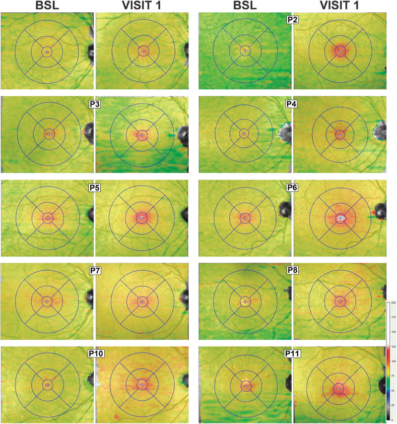
Methods: Patients with stage IV metastatic melanoma and BRAF V600E mutations (n = 11, 31-68 years of age) were included. Treatment was with oral dabrafenib, 150 mg bid, trametinib, 2 mg/day, and HCQ, 400 mg to 600 mg bid. An ophthalmic examination, spectral domain optical coherence tomography, near-infrared and short-wavelength fundus autofluorescence, and static perimetry were performed at baseline, 1 month, and q/6 months after treatment.

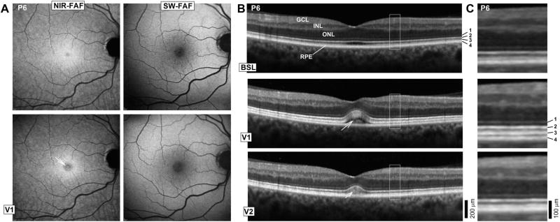
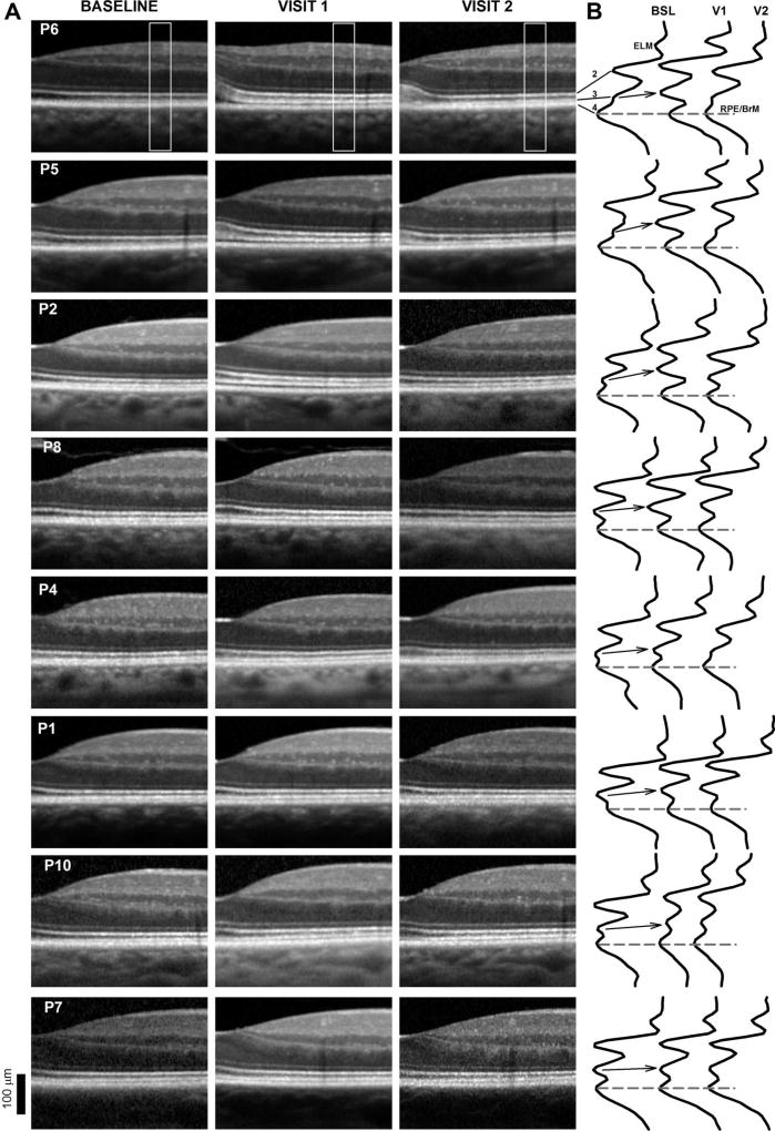
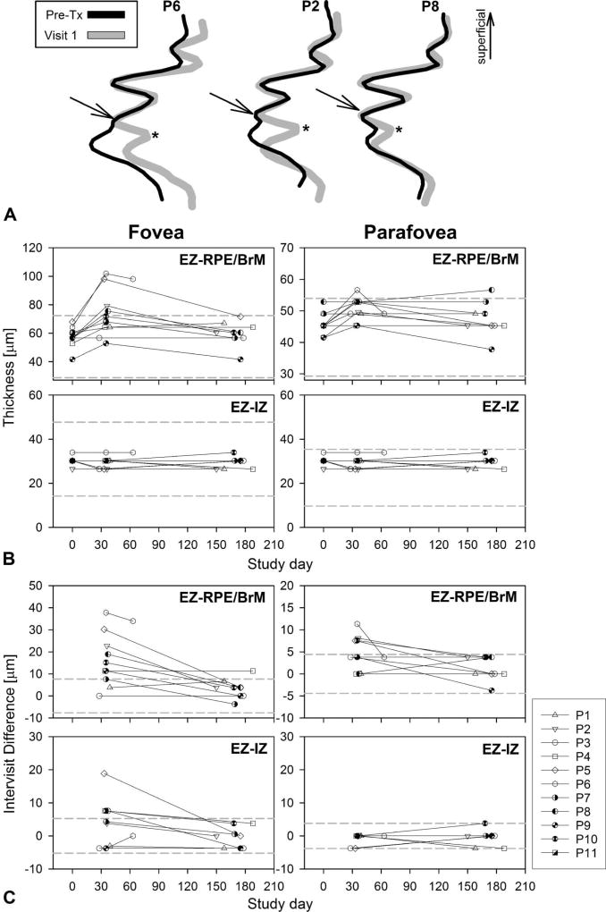
Results: There were no clinically significant ocular events; there was no ocular inflammation. The only medication-related change was a separation of the photoreceptor outer segment tip from the apical retinal pigment epithelium that could be traced from the fovea to the perifoveal retina noted in 9/11 (82%) of the patients. There were no changes in retinal pigment epithelium melanization or lipofuscin content by near-infrared fundus autofluorescence and short-wavelength fundus autofluorescence, respectively. There were no inner retinal or outer nuclear layer changes. Visual acuities and sensitivities were unchanged.

Conclusion: BRAFi (trametinib) + MEKi (dabrafenib) + HCQ causes very frequent, subclinical separation of the photoreceptor outer segment from the apical retinal pigment epithelium without inner retinal changes or signs of inflammation. The changes suggest interference with the maintenance of the outer retinal barrier and/or phagocytic/pump functions of the retinal pigment epithelium by effective MEK inhibition.


Conflict of interest statement

None of the authors has any conflicting interests to disclose.

Figures

Fig. 1
Fig. 1
A. En face retinal imaging of the RPE melanin content using NIR-FAF, and lipofuscin content with SW-FAF in a patient with the most prominent abnormalities. Images were obtained at baseline (BSL) and at a visit (V1), when retinal changes were first noted. Arrow in NIR-FAF V1 points to the foveal center. B. Six millimeter long horizontal SD-OCT cross-sections through the fovea in the patient at BSL, V1, and 6 days (V2) after discontinuation of the study medications because of systemic side effects. Nuclear layers are labeled (outer nuclear layer [ONL]; inner nuclear layer [INL]; ganglion cell layer [GCL]). Outer photoreceptor/RPE laminae are numbered to the side of the BSL image: 1, ELM; 2, EZ; 3, IZ; and 4, RPE/BrM. C. Magnified images corresponding to regions delimited by white squares in (B) show details of the outer photoreceptor and RPE/BrM structure.
Fig. 2
Fig. 2
A. Spectral domain OCT cross-sections from the fovea to 1.4 mm in the nasal retina in 8 of the patients at baseline (BSL), Visit 1 (V1, 30 days posttreatment initiation), and Visit 2 (V2, 180 days posttreatment, 6 days after treatment discontinuation for P6). Patients are ordered from top to bottom by the magnitude of the structural abnormalities. Only the right eye shown for clarity. B. Longitudinal reflectivity profiles obtained from the region area (0.9 mm in the nasal retina) boxed in (A). Longitudinal reflectivity profiles are normalized to the RPE/BrM signal amplitude. Dashed lines connecting RPE/BrM peaks provide a reference; arrows visually connect the location of the IZ signal from baseline to Visit 1 (V1).
Fig. 3
Fig. 3
Topography of the outer retinal changes after MEK/BRAF inhibition. Shown are 30° × 25° topography maps of the distance (or thickness) between the internal limiting membrane and the RPE/BrM layer defined by SD-OCT in 8 of the patients at their baseline (BSL) visits compared with a visit (V1) ~30 days after initiation of the clinical trial medications. Only the right eye shown for clarity. Thickness values (in µm) are mapped to a color scale (bottom right). Superimposed is an early treatment diabetic retinopathy study grid centered at the foveola. Concentric circles of increasing radii divide the central macula in subfields: Central (500 µm radius), inner parafoveal (1,500 µm), and outer perifoveal (3,000 µm).
Fig. 4
Fig. 4
Timecourse of the structural changes. A. Longitudinal reflectivity profiles of the outer retinal sublaminae obtained from the nasal parafovea pretreatment (Pre-TX) are overlaid on LRP from visit 1 (gray traces), ~30 days after medication onset of representative patients. Arrows point to the IZ signal peak as an independent component separated by a trough (asterisk) from the RPE. The traces overlap perfectly in the segment between the EZ and the IZ supporting no changes in POS length. B. Timecourse of the foveal and parafoveal (1.5 mm of eccentricity) changes in thickness for the EZ-to-RPE/BrM and EZ-to-IZ layer distances. The latter relates to the length of the POS. Dashed lines are normal mean ± 2 SD (n = 68, ages 8–61 years). C. Timecourse of the foveal and parafoveal (1.5 mm of eccentricity in the nasal retina) thickness expressed as a difference between the value on a given study date and measurements pretreatment. Dashed lines define mean ± 2 SD of the intervisit variability of the parameter determined at baseline visits and in normal subjects.

References

    1. Flaherty KT, Robert C, Hersey P, et al. Improved survival with MEK inhibition in BRAF-mutated melanoma. N Engl J Med. 2012;367:107–114. - PubMed
    1. Pasquali S, Chiarion-Sileni V, Rossi CR, Mocellin S. Immune checkpoint inhibitors and targeted therapies for metastatic melanoma: a network meta-analysis. Cancer Treat Rev. 2017;54:34–42. - PubMed
    1. Eggermont AM, Spatz A, Robert C. Cutaneous melanoma. Lancet. 2014;383:816–827. - PubMed
    1. Ugurel S, Röhmel J, Ascierto PA, et al. Survival of patients with advanced metastatic melanoma: the impact of novel therapies. Eur J Cancer. 2016;53:125–134. - PubMed
    1. Obaid NM, Bedard K, Huang WY. Strategies for overcoming resistance in tumours harboring BRAF mutations. Int J Mol Sci. 2017;18 doi: 10.3390/ijms18030585. - DOI - PMC - PubMed

Publication types

MeSH terms