Comment
. 2018 Jan 1;141(1):7-10.
doi: 10.1093/brain/awx333.

Early nucleus basalis of Meynert degeneration predicts cognitive decline in Parkinson's disease

Affiliations
Comment

Early nucleus basalis of Meynert degeneration predicts cognitive decline in Parkinson's disease

James P Gratwicke et al. Brain. .

Abstract

This scientific commentary refers to ‘In vivo cholinergic basal forebrain atrophy predicts cognitive decline in de novo Parkinson’s disease’ by Ray et al. (doi:10.1093/brain/awx310).


Figures

Figure 1
Figure 1
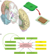
Schematic of anatomy of the nucleus basalis of Meynert (NBM) and risk factors for cognitive decline and dementia in Parkinson’s disease. (A) 3D representation of the anatomy of the NBM. Left: The medial surface of the left hemisphere of the human brain is closest to the viewer. The NBM is the green structure located in the basal forebrain inferior to the globus pallidus internus (GPi). The green arrows emanating from the NBM represent its corticopetal projections passing in the medial and lateral cholinergic pathways to all cortical areas. A = amygdala; C = caudate; P = putamen; T = thalamus. Right: The inset shows a 3D representation of the NBM itself. It is an elongated flat structure spanning 13–14 mm in the sagittal plane. The posterior subsector of the nucleus termed ‘Ch4p’ is highlighted. The brown nucleus below represents an atrophic NBM, reduced in size compared to its previous intact form (represented in translucent green). (B) Model showing all the clinical features and biomarkers which have been shown to be predictive of cognitive decline and future dementia in Parkinson’s disease. Those in green boxes all have a putative cholinergic basis, while those in yellow boxes represent non-cholinergic factors. The green arrows highlight the fact that degeneration of the NBM plays a major role in development of the linked predictive clinical features. Aβ42 = amyloid-β42; DAT = dopamine active transporter scan; REM = rapid eye movement.

Comment on

References

    1. Candy JM, Perry RH, Perry EK, Irving D, Blessed G, Fairbairn AF, et al.Pathological changes in the nucleus of Meynert in Alzheimer’s and Parkinson’s diseases. J Neurol Sci 1983; 59: 277–89. - PubMed
    1. Choi SH, Jung TM, Lee JE, Lee SK, Sohn YH, Lee PH. Volumetric analysis of the substantia innominata in patients with Parkinson’s disease according to cognitive status. Neurobiol Aging 2012; 33: 1265–72. - PubMed
    1. Fling BW, Cohen RG, Mancini M, Nutt JG, Fair DA, Horak FB. Asymmetric pedunculopontine network connectivity in parkinsonian patients with freezing of gait. Brain 2013; 136: 2405–18. - PMC - PubMed
    1. Goldman JG, Williams-Gray C, Barker RA, Duda JE, Galvin JE. The spectrum of cognitive impairment in Lewy body diseases. Mov Disord 2014; 29: 608–21. - PMC - PubMed
    1. Gratwicke J, Jahanshahi M, Foltynie T. Parkinson’s disease dementia: a neural networks perspective. Brain 2015; 138: 1454–76. - PMC - PubMed

Publication types

Substances