. 2018 Apr 1;28(4):223-232.
doi: 10.1093/glycob/cwy002.

A comprehensive Caenorhabditis elegans N-glycan shotgun array

Affiliations

A comprehensive Caenorhabditis elegans N-glycan shotgun array

Ewa Jankowska et al. Glycobiology. .

Abstract

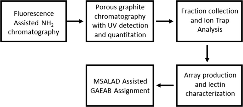
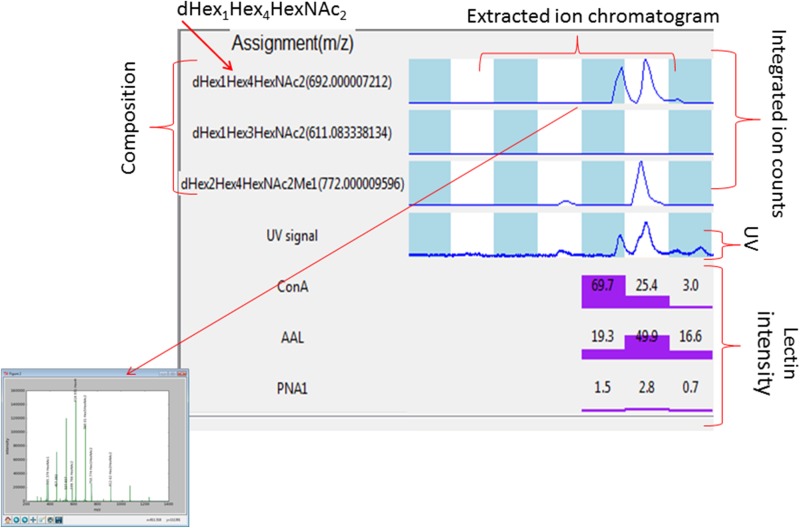
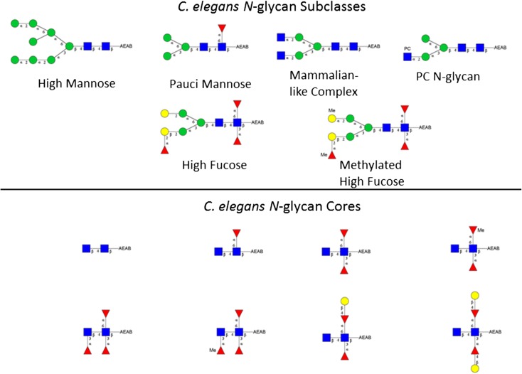
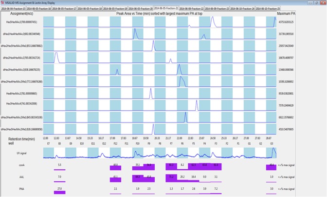
Here we present a Caenorhabditis elegans N-glycan shotgun array. This nematode serves as a model organism for many areas of biology including but not limited to tissue development, host-pathogen interactions, innate immunity, and genetics. Caenorhabditis elegans N-glycans contain structural motifs that are also found in other nematodes as well as trematodes and lepidopteran species. Glycan binding toxins that interact with C. elegans glycoconjugates also do so with some agriculturally relevant species, such as Haemonchus contortus, Ascaris suum, Oesophagostomum dentatum and Trichoplusia ni. This situation implies that protein-carbohydrate interactions seen with C. elegans glycans may also occur in other species with related glycan structures. Therefore, this array may be useful to study these relationships in other nematodes as well as trematode and insect species. The array contains 134 distinct glycomers spanning a wide range of C. elegans N-glycans including the subclasses high mannose, pauci mannose, high fucose, mammalian-like complex and phosphorylcholine substituted forms. The glycans presented on the array have been characterized by two-dimensional separation, ion trap mass spectrometry, and lectin affinity. High fucose glycans were well represented and contain many novel core structures found in C. elegans as well as other species. This array should serve as an investigative platform for carbohydrate binding proteins that interact with N-glycans of C. elegans and over a range of organisms that contain glycan motifs conserved with this nematode.


Figures

Fig. 1.
Fig. 1.
Workflow diagram. Caenorhabditis elegans N-glycans were first separated using a low resolution NH2 column chromatography. Chromatography and quantitation of saccharides was assisted with fluorescence detection. Resultant fractions were individually separated using high resolution porous graphite chromatography in line with ion trap mass spectrometry. Infusion into the mass spectrometer as well as fraction collection was facilitated using a nanomate Triversa instrument. Glycan fractions were quantitated based on photodiode array detected UV absorbance and arrayed accordingly. An array of lectins was used to characterize arrayed GAEABs. Assignment of absorbance, lectin signal and mass spectrometry data were aided with the use of MSALAD and GlycoWorkBench.
Fig. 2.
Fig. 2.
Example MSALAD display and assignment aid. Glycan compositions are assigned using MSALAD monomer database. The software searches datasets for glycans by iteratively matching deconvoluted masses to a structural database. The database iteratively builds glycans based on monomer and substituent sets. Extracted ion chromatographs of assigned glycan compositions are displayed within each LC/MS run. Integrated counts for each composition are shown. Compositions are displayed along with ion m/z from which they were derived. The UV absorbance trace is shown. Point and click displays assigned MS2 mass spectrum. Normalized lectin intensities for Con A, HPA, AAL, PNA and SNA are shown. MSALAD was used as a first pass aid in assignment.
Fig. 3.
Fig. 3.
Representative C. elegans AEAB labeled N-glycans. Subclasses are shown. The upper panel shows high mannose, pauci mannose, PC N-glycan, high fucose and methylated high fucose glycan representatives. The lower panel shows C. elegans core diversity as detected in this study. Designated linkages shown are inferred from the literature and our data.
Fig. 4.
Fig. 4.
MSALAD display of Fraction 21 data. Separation of isomers can be seen for Hex5HexNAc2, dHex1Hex4HexNAc2, dHex2Hex4HexNAc2 and dHex3Hex4HexNAc2. Strong binding of PNA lectin (Gal) is seen for glycomer 28 in well E8. This glycomer has terminally linked Gal on the core.

Similar articles

Cited by

References

    1. Ahmed UK, Maller NC, Iqbal AJ, Al-Riyami L, Harnett W, Raynes JG. 2016. The carbohydrate-linked phosphorylcholine of the parasitic nematode product ES-62 modulates complement activation. J Biol Chem. 291:11939–11953. - PMC - PubMed
    1. Barrows BD, Haslam SM, Bischof LJ, Morris HR, Dell A, Aroian RV. 2007. Resistance to Bacillus thuringiensis toxin in Caenorhabditis elegans from loss of fucose. J Biol Chem. 282:3302–3311. - PubMed
    1. Butschi A, Titz A, Walti MA, Olieric V, Paschinger K, Nobauer K, Guo X, Seeberger PH, Wilson IB, Aebi M et al. . 2010. Caenorhabditis elegans N-glycan core beta-galactoside confers sensitivity towards nematotoxic fungal galectin CGL2. PLoS Pathog. 6:e1000717. - PMC - PubMed
    1. Byres E, Paton AW, Paton JC, Lofling JC, Smith DF, Wilce MC, Talbot UM, Chong DC, Yu H, Huang S et al. . 2008. Incorporation of a non-human glycan mediates human susceptibility to a bacterial toxin. Nature. 456:648–652. - PMC - PubMed
    1. Chen S, Spence AM, Schachter H. 2003. Isolation of null alleles of the Caenorhabditis elegans gly-12, gly-13 and gly-14 genes, all of which encode UDP-GlcNAc: Alpha-3-D-mannoside beta1,2-N-acetylglucosaminyltransferase I activity. Biochimie. 85:391–401. - PubMed

Publication types

MeSH terms

Substances