Review
. 2018 Nov 1;29(13):1237-1272.
doi: 10.1089/ars.2017.7485. Epub 2018 Feb 21.

Redox Paradox: A Novel Approach to Therapeutics-Resistant Cancer

Affiliations
Review

Redox Paradox: A Novel Approach to Therapeutics-Resistant Cancer

Luksana Chaiswing et al. Antioxid Redox Signal. .

Abstract

Significance: Cancer cells that are resistant to radiation and chemotherapy are a major problem limiting the success of cancer therapy. Aggressive cancer cells depend on elevated intracellular levels of reactive oxygen species (ROS) to proliferate, self-renew, and metastasize. As a result, these aggressive cancers maintain high basal levels of ROS compared with normal cells. The prominence of the redox state in cancer cells led us to consider whether increasing the redox state to the condition of oxidative stress could be used as a successful adjuvant therapy for aggressive cancers. Recent Advances: Past attempts using antioxidant compounds to inhibit ROS levels in cancers as redox-based therapy have met with very limited success. However, recent clinical trials using pro-oxidant compounds reveal noteworthy results, which could have a significant impact on the development of strategies for redox-based therapies.

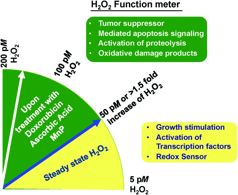
Critical issues: The major objective of this review is to discuss the role of the redox state in aggressive cancers and how to utilize the shift in redox state to improve cancer therapy. We also discuss the paradox of redox state parameters; that is, hydrogen peroxide (H2O2) as the driver molecule for cancer progression as well as a target for cancer treatment.

Future directions: Based on the biological significance of the redox state, we postulate that this system could potentially be used to create a new avenue for targeted therapy, including the potential to incorporate personalized redox therapy for cancer treatment.

Keywords: H2O2; personalized redox therapy; redox state; resistant cancer; rewired redox state.


Figures

<b>FIG. 1.</b>
FIG. 1.
Subcellular redox state and H2O2 concentrations versus cellular functions. (A) Nuclear redox state regulates cancer proliferation. ROS and MnSOD regulate cell cycle, whereas H2O2 and redox thiol couples regulate transcription factors. (B) Mitochondrial redox state regulates cancer metabolism. Mitochondrial ROS and MnSOD regulate glucose consumption, whereas H2O2 and redox thiol couples regulate cancer metabolism via modulation of antioxidants, metabolites, and TCA cycle-associated enzymes. (C) Cytoplasmic redox state regulates cancer growth. Redox thiol couples and low level of H2O2 (nM) act as redox sensors that regulate cellular function through post-translational modification, that is, S-glutathionylation. In contrast, high levels of H2O2M) regulate APs and activate apoptosis via activation of protein adducts. (D) Extracellular redox state regulates cancer metastasis. Redox thiol couples activate receptors-mediated cell growth and cell membrane ROS-generating enzymes. Subsequently, these extracellular ROS activate MMP activities and enhance TGFβ-mediated EMT. Details of how redox thiol couples and H2O2 regulate these targets are provided in text sections. Due to space limitation, several of these targets are not extensively defined. APs, antioxidant proteins; CAT, catalase; Cys, cysteine; CySS, cystine; EMT, epithelial-mesenchymal transition; GPx, glutathione peroxidase; GSH, glutathione; GSSG, glutathione disulfide; H2O2, hydrogen peroxide; HIF-1α, hypoxia inducible factor-1α; Keap1, Kelch-like ECH-associated protein 1; LPO, lipid peroxidation; MnSOD, manganese superoxide dismutase; MMP, matrix metalloproteinase; NO, nitric oxide; Nrf2, nuclear factor-erythroid 2-related factor 2; O2•−, superoxide radical; ONOO, peroxinitrite; Prx, peroxiredoxin; ROS, reactive oxygen species; STAT3, signal transducer and activator of transcription factor 3; Trx, thioredoxin. To see this illustration in color, the reader is referred to the web version of this article at www.liebertpub.com/ars
<b>FIG. 2.</b>
FIG. 2.
Increased ROS level as an anticancer therapy approach to treat aggressive cancers with adapted rewired redox state. Cancer cells are usually under high oxidizing conditions (pink range) due to rewired redox state process (often due to increased influxes of ROS/RNS). Adaptation to persistent and high levels of ROS can promote metastasis and resistance of cancers. However, shifting redox state to an extreme oxidizing condition will push cancer cells into the death zone (blue area). Since aggressive cancers, including radioresistant, chemoresistant, and metastasis cancers, rewired their redox state to an oxidized status higher than that of their parental cancers, using an ROS-generating drug to push redox state into the death zone (brown arrow) seems appropriate. In normal cells, cellular redox status is kept at a low oxidizing level compared with a cancerous condition (yellow range). A small shift in cellular redox status toward an oxidizing condition will stimulate adaptive signaling, which leads to upregulation of the APs system and promotes normal cell survival. Generally, the redox potential range is −150 to −300 mV, whereas H2O2 concentration range is 0 to ∼100 nM. RNS, reactive nitrogen species. To see this illustration in color, the reader is referred to the web version of this article at www.liebertpub.com/ars
<b>FIG. 3.</b>
FIG. 3.
Increased levels of oxidative stress markers, 4HNE and PrxSO3 in aggressive prostate cancer. Quantitative analysis of immunohistochemistry staining of 4HNE and PrxSO3 expression from tumor microarrays of prostate cancer patients using the Aperio system. (A) Tissues from metastatic prostate cancer stage 4 (Metastasis_S4). (B) Tissues from patients who died from prostate cancer. Deceased_S4 = PCa who died from stage 4 PCa. *p < 0.05, #p = 0.08. y-axis = Pixel intensity of oxidative stress marker staining. 4HNE, 4-hydroxynonenal; PrxSO3, oxidation form of Prx. To see this illustration in color, the reader is referred to the web version of this article at www.liebertpub.com/ars
<b>FIG. 4.</b>
FIG. 4.
Proposed models of therapeutic-resistant cancers. Accumulated evidence demonstrates that therapeutics-resistant cancers, such as radioresistant cancer, often correlate with (i) stem cell markers, (ii) high OXPHOS gene expression and activity, (iii) shift of energetic status, and (iv) rewired redox state features (high ROS, oxidized redox status, alteration of APs). These features are the foundation of a well-tolerated cancer phenotype. Two proposed models explain potential recurrence of cancers after standard treatments. (A) Pre-existing therapeutic resistance/recurrence cancers. In this model, the recurrent cancer features, including rewired redox state, exist before the treatment. Therefore, cancer cells that exhibit therapeutics-resistant features escape the hostile environment of the treatment and progress into a more advanced therapeutic-resistant cancer (Red cells). (B) Post-treatment therapeutic-resistant/recurrent cancers. In this model, cancer cells demonstrate no indication of therapeutics-resistant features. However, after the standard treatments, survival cancer cells develop diverse adaptive mechanisms, including rewired redox state, that are able to avoid the hostile environment. Subsequently, these cells develop into recurrent cancers (dark blue cells). OXPHOS, oxidative phosphorylation. To see this illustration in color, the reader is referred to the web version of this article at www.liebertpub.com/ars
<b>FIG. 5.</b>
FIG. 5.
The interplay between concentrations and functions of H2O2 in cancer cells. H2O2 is a potential candidate for a key molecule that decides the fate of cancer survival. Based on 3-AT-mediated inactivation of CAT assay, the steady state of H2O2 in selected cancer cells ranges from 5 to 50 pM (4, 69, 85, 253, 348). This range is associated with the following cancer responses: redox sensor on proteins, growth stimulation, and activation of transcription factors. In contrast, influxes that increase H2O2 concentrations more than 1.5-fold of its steady state via treatment of ROS-generating drugs, such as doxorubicin, AA, and MnP, lead to oxidatively damaged proteins, activation of proteolysis, mediation of apoptosis, and cancer cell death. 3-AT, 3-aminotriazole; AA, ascorbic acid; MnP, Mn(III) meso-tetrakis (N-n-butoxyethyl-pyridinium-2yl) porphyrin; pM, picomolar. To see this illustration in color, the reader is referred to the web version of this article at www.liebertpub.com/ars
<b>FIG. 6.</b>
FIG. 6.
Proposed mechanism of how mitochondrial pro-oxidant compounds mediate death in platinum-resistant cancers. Treatment with platinum drugs (cisplatin/carboplatin) activates DNA adducts that induce DNA damage in the nucleus. Many resistant mechanisms are observed in cancers that are resistant to platinum treatment, such as drug efflux, poor accessibility, and detoxification enzymes. One resistant mechanism is the formation of platinum-GSH conjugations, especially in cancers that often maintain a high level of GSH due to rewired redox state. Mitochondrial pro-oxidant agents such as MnP induce the following mechanisms: (1) Mitochondrial ROS generation. Influx of mitochondrial ROS inhibits mitochondrial function and activates mitochondrial-dependent apoptosis via BAX/Cytochrome C pathways. (2) Cellular ROS/RNS production. Induction of cellular oxidative stress utilizes GSH and prevents platinum-GSH conjugations. (3) MnP-GSH conjugations. MnP directly or indirectly binds with GSH, prevents platinum drug conjugations, and releases drugs that induce DNA damage. Therefore, co-treatment with mitochondrial pro-oxidant compounds such as MnP would instantaneously induce DNA damage and mitochondrial stress, which negatively affect the capacity of cancer cells to respond simultaneously to oxidative stress in multiple subcellular compartment sites. MRP1, multi-drug resistance-associated protein 1. To see this illustration in color, the reader is referred to the web version of this article at www.liebertpub.com/ars
<b>FIG. 7.</b>
FIG. 7.
RNA expression profiles of candidates for redox state index across cancer types. RNA expression of (i) antioxidant proteins. CAT and Nrf2, and (ii) ROS-related generating proteins, NOX4, and transferrin receptor, in high prevalence cancers. CAT, Nrf2, NOX4, and transferrin receptor are overexpressed in most cancers except AML, which exhibits low expression of NOX4. Data presented as median. The y-axis = RNA Seq of each protein (Log), in which 0 is a baseline. The x-axis is types of cancers. Red = Missense, Blue = No mutation. AML, acute myeloid leukemia; GBM, gliobastoma; PCPG, pheochromocytoma and paraganglioma. To see this illustration in color, the reader is referred to the web version of this article at www.liebertpub.com/ars
<b>FIG. 8.</b>
FIG. 8.
The potential application of personalized redox therapy for diagnosis, treatment, and surveillance of cancer patients. Accumulated evidence indicates that when an AML patient expresses resistance to cytarabine (AraC), the cause is likely determined by pre-existing and persisting redox, metabolic, and energetic status, independent of stem cell features (91). These patients often exhibit an enriched CD36-FA (fatty acid) translocase receptor expression (a robust biomarker of residual disease), high OXPHOS gene signature, high ROS levels with modified intracellular redox status, and an increase in fatty acid oxidation. To improve the efficacy of AraC for AML patients with established pre-existing resistant/recurrent cancer features, a redox state index that is integrated with other molecular biomarkers is proposed. Note that all biomarkers and proposed strategies are potential targets, which requires evaluation in well-designed clinical trials. CPT1, carnitine O-palmitoyltransferase 1; TFAM, mitochondrial transcription factor A. To see this illustration in color, the reader is referred to the web version of this article at www.liebertpub.com/ars

References

    1. Abdel-Latif MM, Raouf AA, Sabra K, Kelleher D, and Reynolds JV. Vitamin C enhances chemosensitization of esophageal cancer cells in vitro. J Chemother 17: 539–549, 2005 - PubMed
    1. Acharya A, Das I, Chandhok D, and Saha T. Redox regulation in cancer: a double-edged sword with therapeutic potential. Oxid Med Cell Longev 3: 23–34, 2010 - PMC - PubMed
    1. Adimora NJ, Jones DP, and Kemp ML. A model of redox kinetics implicates the thiol proteome in cellular hydrogen peroxide responses. Antioxid Redox Signal 13: 731–743, 2010 - PMC - PubMed
    1. Ahmad IM, Aykin-Burns N, Sim JE, Walsh SA, Higashikubo R, Buettner GR, Venkataraman S, Mackey MA, Flanagan SW, Oberley LW, and Spitz DR. Mitochondrial O2*- and H2O2 mediate glucose deprivation-induced stress in human cancer cells. J Biol Chem 280: 4254–4263, 2005 - PubMed
    1. Ahmad KA, Clement MV, Hanif IM, and Pervaiz S. Resveratrol inhibits drug-induced apoptosis in human leukemia cells by creating an intracellular milieu nonpermissive for death execution. Cancer Res 64: 1452–1459, 2004 - PubMed

Publication types

Substances

LinkOut - more resources