

Late normal tissue response in the rat spinal cord after carbon ion irradiation
- PMID: 29325596
- PMCID: PMC5765675
- DOI: 10.1186/s13014-017-0950-5
Late normal tissue response in the rat spinal cord after carbon ion irradiation
Abstract
Background: The present work summarizes the research activities on radiation-induced late effects in the rat spinal cord carried out within the "clinical research group ion beam therapy" funded by the German Research Foundation (DFG, KFO 214).
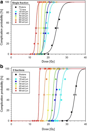
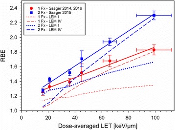
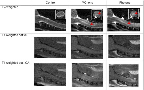
Methods and materials: Dose-response curves for the endpoint radiation-induced myelopathy were determined at 6 different positions (LET 16-99 keV/μm) within a 6 cm spread-out Bragg peak using either 1, 2 or 6 fractions of carbon ions. Based on the tolerance dose TD50 of carbon ions and photons, the relative biological effectiveness (RBE) was determined and compared with predictions of the local effect model (LEM I and IV). Within a longitudinal magnetic resonance imaging (MRI)-based study the temporal development of radiation-induced changes in the spinal cord was characterized. To test the protective potential of the ACE (angiotensin converting enzyme)-inhibitor ramipril™, an additional dose-response experiment was performed.
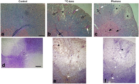
Results: The RBE-values increased with LET and the increase was found to be larger for smaller fractional doses. Benchmarking the RBE-values as predicted by LEM I and LEM IV with the measured data revealed that LEM IV is more accurate in the high-LET, while LEM I is more accurate in the low-LET region. Characterization of the temporal development of radiation-induced changes with MRI demonstrated a shorter latency time for carbon ions, reflected on the histological level by an increased vessel perforation after carbon ion as compared to photon irradiations. For the ACE-inhibitor ramipril™, a mitigative rather than protective effect was found.
Conclusions: This comprehensive study established a large and consistent RBE data base for late effects in the rat spinal cord after carbon ion irradiation which will be further extended in ongoing studies. Using MRI, an extensive characterization of the temporal development of radiation-induced alterations was obtained. The reduced latency time for carbon ions is expected to originate from a dynamic interaction of various complex pathological processes. A dominant observation after carbon ion irradiation was an increase in vessel perforation preferentially in the white matter. To enable a targeted pharmacological intervention more details of the molecular pathways, responsible for the development of radiation-induced myelopathy are required.
Keywords: Carbon ion therapy; Magnetic resonance imaging; Radiation induced myelopathy; Rat spinal cord; Relative biological effectiveness (RBE).
Conflict of interest statement
Ethics approval
All experiments were approved by the governmental review committee on animal care (35–9185.81/G62–08, G117/13, G34/13).
Consent for publication
Not applicable.
Competing interests
The authors declare that they have no competing interests.
Publisher’s Note
Springer Nature remains neutral with regard to jurisdictional claims in published maps and institutional affiliations.
Figures
References
-
- Uhl M, Herfarth K, Debus J. Comparing the use of protons and carbon ions for treatment. Cancer J. 2014;20(6):433–439. - PubMed
-
- Jensen AD, Nikoghosyan AV, Poulakis M, et al. Combined intensity-modulated radiotherapy plus raster-scanned carbon ion boost for advanced adenoid cystic carcinoma of the head and neck results in superior locoregional control and overall survival. Cancer. 2015;121(17):3001–3009. - PubMed
-
- Uhl M, Mattke M, Welzel T, et al. High control rate in patients with chondrosarcoma of the skull base after carbon ion therapy: first report of long-term results. Cancer. 2014;120(10):1579–1585. - PubMed
-
- Uhl M, Mattke M, Welzel T, et al. Highly effective treatment of skull base chordoma with carbon ion irradiation using a raster scan technique in 155 patients: first long-term results. Cancer. 2014;120(21):3410–3417. - PubMed
MeSH terms
Substances
Grants and funding
LinkOut - more resources
Full Text Sources
Other Literature Sources
Miscellaneous
