Review
. 2018 May 18;18(2):71-87.
doi: 10.3727/105221618X15156018385515. Epub 2018 Jan 11.

Bile Acid Metabolism in Liver Pathobiology

Affiliations
Review

Bile Acid Metabolism in Liver Pathobiology

John Y L Chiang et al. Gene Expr. .

Abstract

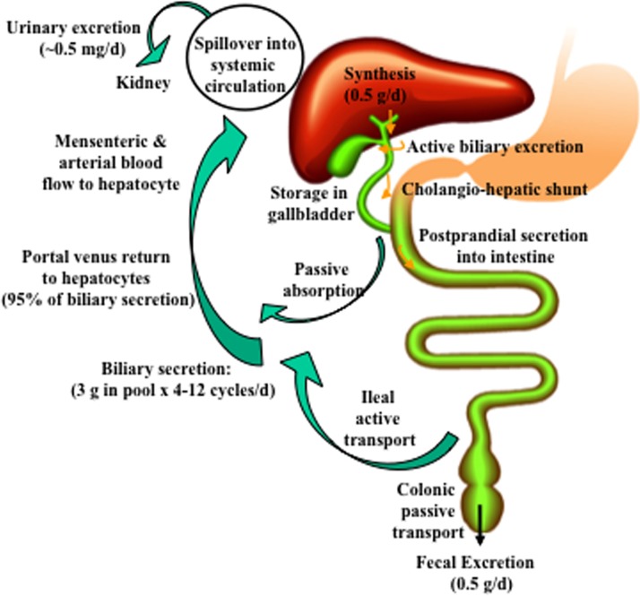
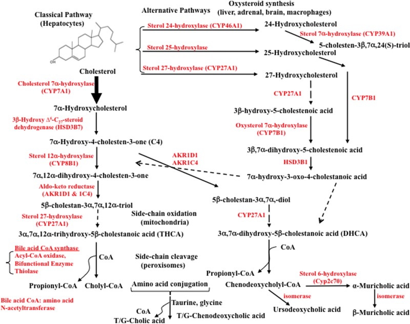
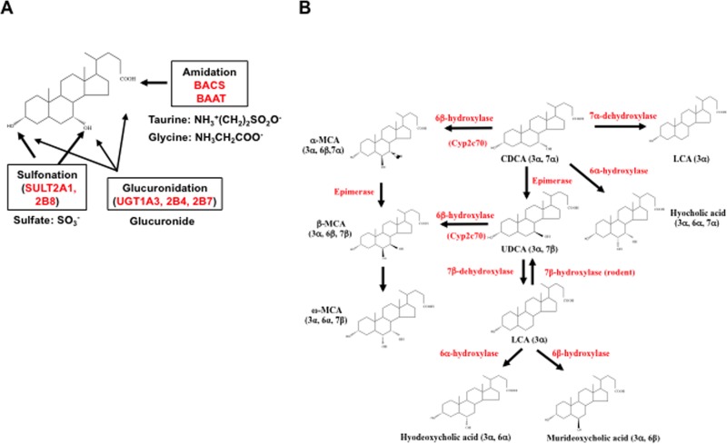
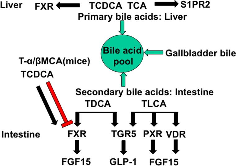
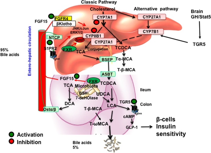
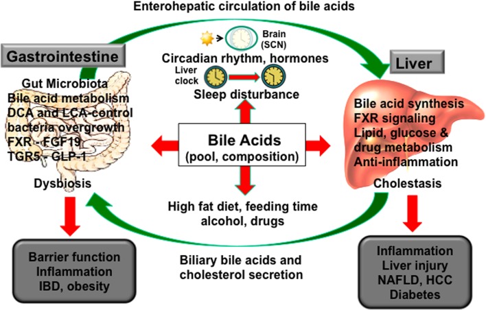
Bile acids facilitate intestinal nutrient absorption and biliary cholesterol secretion to maintain bile acid homeostasis, which is essential for protecting liver and other tissues and cells from cholesterol and bile acid toxicity. Bile acid metabolism is tightly regulated by bile acid synthesis in the liver and bile acid biotransformation in the intestine. Bile acids are endogenous ligands that activate a complex network of nuclear receptor farnesoid X receptor and membrane G protein-coupled bile acid receptor-1 to regulate hepatic lipid and glucose metabolic homeostasis and energy metabolism. The gut-to-liver axis plays a critical role in the regulation of enterohepatic circulation of bile acids, bile acid pool size, and bile acid composition. Bile acids control gut bacteria overgrowth, and gut bacteria metabolize bile acids to regulate host metabolism. Alteration of bile acid metabolism by high-fat diets, sleep disruption, alcohol, and drugs reshapes gut microbiome and causes dysbiosis, obesity, and metabolic disorders. Gender differences in bile acid metabolism, FXR signaling, and gut microbiota have been linked to higher prevalence of fatty liver disease and hepatocellular carcinoma in males. Alteration of bile acid homeostasis contributes to cholestatic liver diseases, inflammatory diseases in the digestive system, obesity, and diabetes. Bile acid-activated receptors are potential therapeutic targets for developing drugs to treat metabolic disorders.


Conflict of interest statement

The authors declare no conflicts of interests.

Figures

Figure 1
Figure 1
Enterohepatic circulation of bile acids. About 0.5 g of bile acids is synthesized per day in an average adult. Bile acids are secreted into bile and stored in the gallbladder. Small amounts of bile acids are circulated from cholangiocytes to the liver via the cholangio-hepatic shunt. After each meal, bile acids are excreted to the intestinal tract. A small amount of bile acids is passively absorbed in the upper intestine into mesenteric and arterial blood flow to hepatocytes. Most bile acids are reabsorbed in the ileum by active transport via apical sodium-dependent bile salt transporter (ASBT) and transported back to the liver via portal blood circulation. A constant bile acid pool (3 g) is circulated 4 to 12 times a day. Approximately 95% of bile acids in bile are recirculated back to the liver, and about 5% (0.5 g) lost in fecal excretion are replenished by de novo synthesis in the liver. A small amount (0.5 mg/day) of bile acid spillover into systemic circulation is cleared in urine.
Figure 2
Figure 2
Bile acid synthesis pathways. In the liver, cholesterol 7α-hydroxylase (CYP7A1) initiates the classical bile acid synthesis pathway by hydroxylation of the steroid rings at 7α-C for further modifications of the steroid rings, followed by steroid side chain oxidation and cleavage, whereas sterol 27-hydroxylase (CYP27A1) initiates the alternative bile acid synthesis pathway by oxidation of the steroid side chain followed by modifications of the steroid rings and cleavage of the side chain in the classic pathway. CYP27A1 is expressed in most tissues and macrophages. Sterol 25-hydroxylase (a non-CYP450 enzyme) in the liver and steroid 24-hydroxylase (CYP46A1) in the brain also oxidize cholesterol. In the alternative pathways, a nonspecific oxysterol 7α-hydroxylase (CYP7B1) hydroxylates 27-hydroxycholesterol and 25-hydroxycholesterol, whereas a specific sterol 7α-hydroxylase (CYP39A1) hydroxylates 24-hydroxycholesterol. Only the liver has all the enzymes required for the synthesis of cholic acid (CA) and chenodeoxycholic acid (CDCA), the two primary bile acids synthesized in humans (shown on the left, see text for details). The oxidized steroid intermediates (oxysterols) produced in the extrahepatic tissues can be used for bile acid synthesis in the liver. Sterol 12α-hydroxylase (CYP8B1) is required for CA synthesis. Without 12α-hydroxylation, CDCA is synthesized. Following steroid side chain cleavage, cholyl-CoA and chenodeoxycholyl-CoA are conjugated to amino acids, either taurine or glycine. In mice, CDCA is 6α-hydroxylated to form α-muricholic acid (α-MCA) by a sterol-6α-hydroxylase (Cyp2c70) catalyzed reaction. The 7α-OH group in α-MCA is epimerized (isomerized) to a 7β-hydroxyl group to form β-MCA. The 7α-HO group in CDCA can be epimerized to 7β-HO to form ursodeoxycholic acid (UDCA), a highly soluble bile acid in humans and mice.
Figure 3
Figure 3
Bile acid conjugation and transformation. (A) Bile acid conjugation. In addition to conjugation to taurine and glycine at the C24-carboxyl group by bile acid Co-A synthase (BACS) and bile acid CoA: amino acid N-acetyltransferase (BAAT), bile acids can be conjugated to sulfate by bile acid sulfotransferase (SULT2A1, SULT2B8) at the C3 and C7 positions, or glucuronidated by UDP-glucuronosyl transferase (UGT1A3, 2B4, and 2B7). (B) Bile acid transformation in enterocytes. Primary bile acids, CDCA, CA, and UDCA, can be converted to other bile acids in the liver and intestine. In the liver, CDCA can be converted to α-MCA, β-MCA, and UDCA as described in Figure 2. In the colon, bacterial bile salt hydratases (dehydroxylases) first deconjugate bile acids to free bile acids, and then gut bacteria converts CA to deoxycholic acid (DCA). CDCA can be converted to lithocholic acid (LCA) by 7α-dehydroxylase and hyocholic acid by 6α-hydroxylase. LCA can be converted to hyodeoxycholic acid by 6α-hydroxylase, or murideoxycholic acid by 6β-hydroxylase. In rodents, LCA can be converted to UDCA by 7β-hydroxylase and UDCA can be converted to CDCA. In the colon, α-MCA and β-MCA can be converted to ω-MCA for fecal excretion.
Figure 4
Figure 4
Bile acid-activated receptors in the gastrointestinal system. Bile acid-activated receptors are differentially activated by primary and secondary bile acids in the liver and intestine. The bile acid pool consists both of the primary bile acids TCDCA, TCA, Tα-MCA, and Tβ-MCA, and the secondary bile acids TDCA and TLCA. In the liver, TCDCA activates FXR, whereas TCA activates S1PR2. In the intestine, TCDCA and TDCA activate FXR, whereas Tα-MCA and Tβ-MCA antagonize FXR. TLCA and TDCA activate TGR5, and TLCA activates VDR and PXR.
Figure 5
Figure 5
Mechanisms of bile acid regulation of bile acid synthesis. In the liver, activation of FXR by agonists induces SHP to inhibit CYP7A1 and CYP8B1 gene transcription. TCA activates S1PR2 and may inhibit CYP7A1 via the ERK1/2 pathway. In the intestine, TCDCA activates FXR to induce FGF15, which is transported to hepatocytes to activate FGFR4/β-Klotho receptor, which stimulates the JNK/cJun and ERK1/2 pathways to inhibit CYP7A1 and CYP8B1. In the brain, growth hormone (GH) activates STAT5 signaling to activate TGR5 signaling to induce CYP7B1 in male mice and regulate the alternative bile acid synthesis pathway. In the intestine, TLCA activates TGR5 in L cells to stimulate GLP-1 secretion, which increases insulin sensitivity by stimulating insulin secretion from pancreatic β-cells.
Figure 6
Figure 6
The gut-to-liver axis and circadian rhythms in bile acid metabolism. Bile acid synthesis in the liver is controlled in part by circadian expression of CYP7A1 expression. Bile acids control the gut microbiota population, and gut bacteria regulate bile acid metabolism, bile acid composition, and enterohepatic circulation of bile acids. Sleep disruption, high-fat diet (HFD), alcohol, and drugs alter the central clock in the hypothalamic suprachiasmatic nucleus (SCN) of the brain to desynchronize the peripheral clocks in the liver and intestine. Disruption of circadian rhythms alters bile acid homeostasis, causes liver and intestine inflammation and dysbiosis, and contributes to cholestatic liver injury, nonalcoholic fatty liver disease (NAFLD), diabetes, and inflammatory bowel diseases.

References

    1. Russell DW. Fifty years of advances in bile acid synthesis and metabolism. J Lipid Res. 2009;50(Suppl):S120–5. - PMC - PubMed
    1. Chiang JY. Bile acids: Regulation of synthesis. J Lipid Res. 2009;50:1955–66. - PMC - PubMed
    1. Lefebvre P, Cariou B, Lien F, Kuipers F, Staels B. Role of bile acids and bile acid receptors in metabolic regulation. Physiol Rev. 2009;89(1):147–91. - PubMed
    1. Chiang JY. Bile acid metabolism and signaling. Compr Physiol. 2013;3:1191–212. - PMC - PubMed
    1. Thomas C, Auwerx J, Schoonjans K. Bile acids and the membrane bile acid receptor TGR5—Connecting nutrition and metabolism. Thyroid 2008;18(2):167–74. - PubMed

Publication types

Substances