. 2018 Jan 12;359(6372):232-236.
doi: 10.1126/science.aao1503.

CX3CR1+ mononuclear phagocytes control immunity to intestinal fungi

Affiliations

CX3CR1+ mononuclear phagocytes control immunity to intestinal fungi

Irina Leonardi et al. Science. .

Abstract

Intestinal fungi are an important component of the microbiota, and recent studies have unveiled their potential in modulating host immune homeostasis and inflammatory disease. Nonetheless, the mechanisms governing immunity to gut fungal communities (mycobiota) remain unknown. We identified CX3CR1+ mononuclear phagocytes (MNPs) as being essential for the initiation of innate and adaptive immune responses to intestinal fungi. CX3CR1+ MNPs express antifungal receptors and activate antifungal responses in a Syk-dependent manner. Genetic ablation of CX3CR1+ MNPs in mice led to changes in gut fungal communities and to severe colitis that was rescued by antifungal treatment. In Crohn's disease patients, a missense mutation in the gene encoding CX3CR1 was identified and found to be associated with impaired antifungal responses. These results unravel a role of CX3CR1+ MNPs in mediating interactions between intestinal mycobiota and host immunity at steady state and during inflammatory disease.


Figures

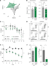
Fig. 1
Fig. 1. CX3CR1 mononuclear cells express antifungal receptors and recognize fungi in the intestine
(A) RNA sequencing (RNA-seq) analysis was performed on sorted CD11b+ CD103+ dendritic cells and CX3CR1+ mononuclear phagocytes. Volcano plot of P-value versus FC comparing gene expression in the two cell subsets; red dots indicate an FDR of <0.05. (B) Logarithmic count per million (log(cpm)) normalization of genes involved in antigen presentation (left panel) or fungal recognition (right panel). (C) The expression of antifungal CLRs was confirmed by RT-qPCR. (D) Representative flow cytometry histogram of dectin-1, dectin-2 and Syk expression among CD11b CD103+; CD11b+ CD103+ and CD11b+ CX3CR1+ cells in colons of WT mice. (E) Representative confocal imaging of the intake of C. albicans-RFP (red) by CX3CR1+ MNPs (green, CX3CR1+, DAPI+) and other cell types (blue, CX3CR1, DAPI+) in the intestine. Bar graphs represent mean ± SEM of individual mice (N=4-7), representative of at least two independent experiments. *P<0.05, **P<0.01, one-way ANOVA.
Fig. 2
Fig. 2. CX3CR1+ MNPs control gut antifungal immunity
Colonic lamina propria cells were collected from ΔIrf4 mice (orange bars) or littermates (lit, grey bars) fed (C.a) or not (NT) with 5.107 C. albicans at day 10. (A and B) Expression of RORγt and IL-17 by CD4+ T cells (pooled from two independent experiments). Cd11c-Cre+/− CX3CR1DTR mice (ΔCX3CR1, green bars) or Cd11c-Cre−/− CX3CR1DTR littermates (lit, grey bars) were treated with diphtheria toxin (DT), and fed with C. albicans. (C and D) RORγt and IL-17 expression by the CD4+ T cells in the colon (pooled from three independent experiments). (E) IgG against the commensal C. tropicalis and flagellin. (F) Systemic IgG responses following i.p injection with C. albicans at day 1, 2 and 5 (pooled from two independent experiments). (G) C. albicans in the feces of control, ΔIrf4 and ΔCX3CR1 mice at day 10 (dots represent individual mice). ΔCX3CR1 mice and littermates were transferred with purified CD4+ Thy1.1+ OT-II cells, fed C. albicans-OVA and sacrificed after 10 days. (H) Representative plots of CD4+ Thy1.1+ OT-II cells proliferation in the colon (pooled from three independent experiments). Cx3cr1-Cre-ERT+/− Sykfl/fl mice (ΔSyk) or littermates (Litt) treated with tamoxifen and fed with C. albicans. (I) RORγt expression by CD4+ T cells in the colon (pooled from two independent experiments). (J) C. albicans in the feces at day 10 (dots represent individual mice). (K) IgG responses against C. tropicalis at day 10 (n=5 per group). (L) Quantification of proliferating CFSE CD4+ Thy1.1+ OT-II cells in the colon (pooled from two independent experiments). Statistical analysis: Data are presented as mean +/− SEM; *P<0.05, **P<0.01, ***P<0.001 (Mann-Whitney Test (E, G, J, K, L), one-way ANOVA (B, C, D, F)).
Fig. 3
Fig. 3. Depletion of CX3CR1+ MNP affects gut mycobiota and results in exacerbated intestinal disease
Feces from ΔCX3CR1 or WT littermates (Litt) mice were collected 7 days after administration of the first DT dose. (A) NMDS plot of distance ordination based on Bray-Curtis dissimilarities in the colon for fungal OTUs (WT n=5; ΔCX3CR1 n=6). (B) Alpha diversity (Simpson diversity index) among the Ascomycota (left) and Basidiomycota (right) phyla; pooled from two independent experiments, mean +/− SEM, each circle denotes one mouse. (C) Weight change during DSS colitis in ΔCX3CR1 or control littermates following short-term treatment with fluconazole (Fl) or no treatment (NT) (pooled from two independent experiments). (D) Representative plots of neutrophil infiltration (CD11b+ Gr-1high) in the colon following DSS administration. (E) Weight change during DSS colitis in ΔCX3CR1 or control littermates fed with C. tropicalis (C.t) (mean +/− SEM, lit n=5; ΔCX3CR1 n=5). (F) C. tropicalis cfu/g in the feces of ΔCX3CR1 or control littermates at day 7 (dots represent individual mice, mean +/− SEM). (K) Systemic IgG responses against C. tropicalis were assessed by ELISA. Statistical analysis: *P<0.05, **P<0.01, ***P<0.001 (Mann-Whitney Test (B, F), two-way ANOVA (C, E)).
Fig. 4
Fig. 4. Polymorphisms in the coding region of the CX3CR1 gene is associated with decreased anti-fungal IgG responses in CD patients
(A) Representative pictures of the intake of fungal species (colored) by CX3CR1+ MNPs (grey) in the colon. (B) Association between the missense mutation rs3732378 and the systemic serologic markers anti-neutrophil cytoplasmic antibodies (anca), flagellin (cbir), Pseudomonas fluorescens – associated sequence I-2 (i2), and anti S. cerevisiae IgG antibodies (igg.asca) among 503 CD patients. FA, frequency affected; FU, frequency unaffected; L95 and U95, lower and upper 95th confidence interval. (C) IgG ASCA and anti-flagellin (cbir) IgG responses were assessed in the sera from rs3732378 homozygous (AA), heterozygous (AG) and control (GG) CD patients by ELISA (dots represent individual patients, bar represent mean). (D) IgG responses against different commensal fungi (Candida albicans, Pichia kudrazevii, Saccharomyces cerevisiae, Aspergillus amstellodamii, Wallemia sebi, Malassezia restricta were assessed (dots represent individual patients, bar represent mean). Statistical analysis: *P<0.05, **P<0.01, ***P<0.001 (Mann-Whitney test (D), one-way ANOVA (C)).

References

    1. Scupham AJ, Presley LL, Wei B, Bent E, Griffith N, McPherson M, Zhu F, Oluwadara O, Rao N, Braun J, Borneman J. Abundant and diverse fungal microbiota in the murine intestine. Appl Environ Microbiol. 2006;72:793–801. - PMC - PubMed
    1. Iliev ID, Leonardi I. Fungal dysbiosis: immunity and interactions at mucosal barriers. Nat Rev Immunol. 2017;17:635–646. - PMC - PubMed
    1. Iliev ID, Funari VA, Taylor KD, Nguyen Q, Reyes CN, Strom SP, Brown J, Becker CA, Fleshner PR, Dubinsky M, Rotter JI, Wang HL, McGovern DP, Brown GD, Underhill DM. Interactions between commensal fungi and the C-type lectin receptor Dectin-1 influence colitis. Science. 2012;336:1314–1317. - PMC - PubMed
    1. Sokol H, Leducq V, Aschard H, Pham HP, Jegou S, Landman C, Cohen D, Liguori G, Bourrier A, Nion-Larmurier I, Cosnes J, Seksik P, Langella P, Skurnik D, Richard ML, Beaugerie L. Fungal microbiota dysbiosis in IBD. Gut. 2017;66:1039–1048. - PMC - PubMed
    1. Wheeler ML, Limon JJ, Bar AS, Leal CA, Gargus M, Tang J, Brown J, Funari VA, Wang HL, Crother TR, Arditi M, Underhill DM, Iliev ID. Immunological Consequences of Intestinal Fungal Dysbiosis. Cell Host Microbe. 2016;19:865–873. - PMC - PubMed

Publication types

MeSH terms