. 2017 Dec 20:8:2555.
doi: 10.3389/fmicb.2017.02555. eCollection 2017.

The Mitochondrial GTPase Gem1 Contributes to the Cell Wall Stress Response and Invasive Growth of Candida albicans

Affiliations

The Mitochondrial GTPase Gem1 Contributes to the Cell Wall Stress Response and Invasive Growth of Candida albicans

Barbara Koch et al. Front Microbiol. .

Abstract

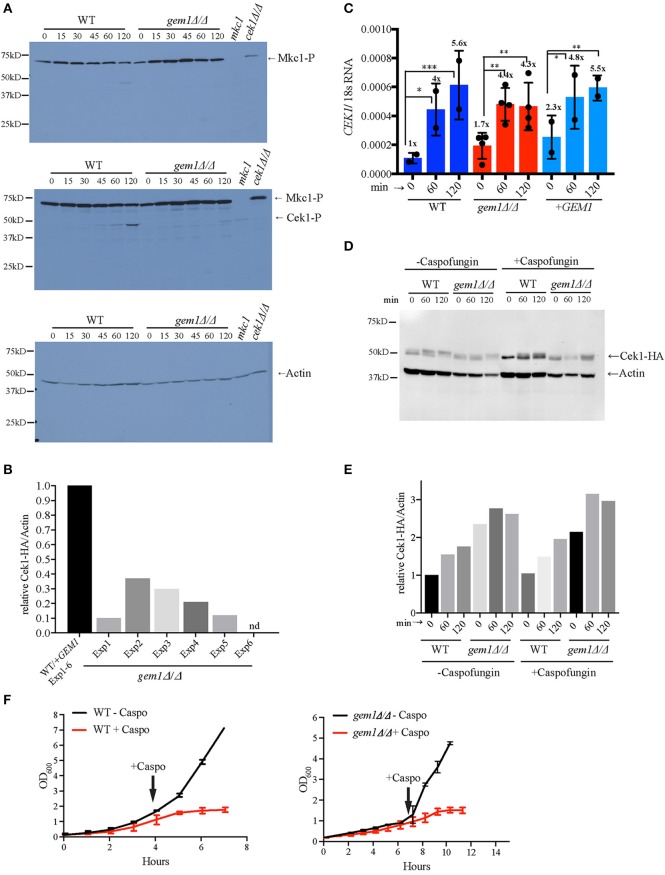
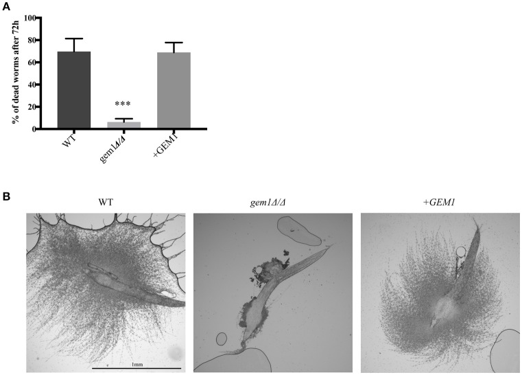
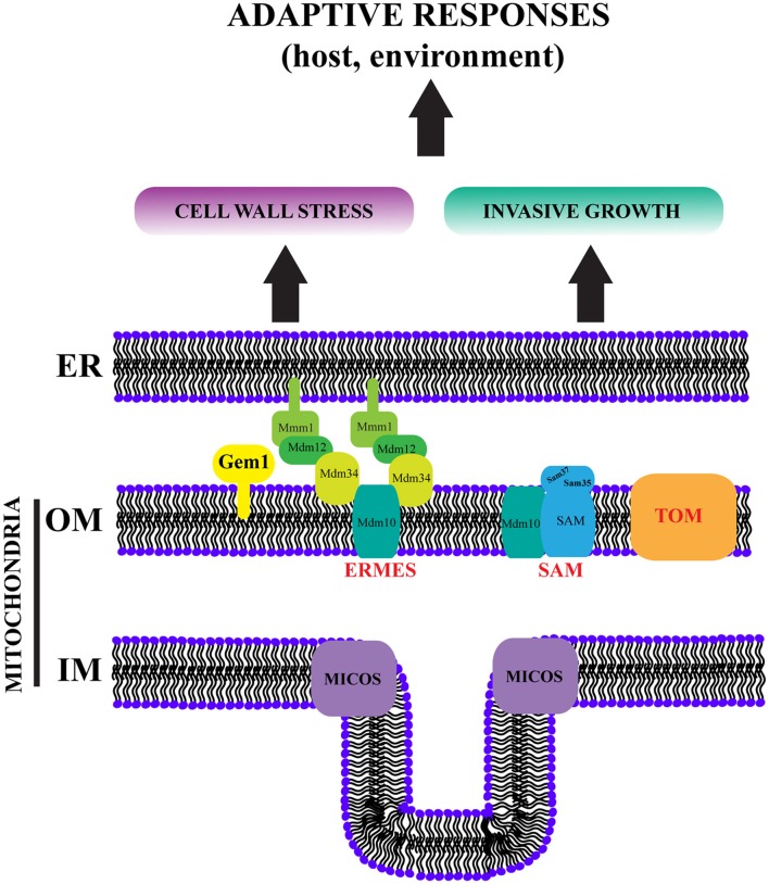
The interactions of mitochondria with the endoplasmic reticulum (ER) are crucial for maintaining proper mitochondrial morphology, function and dynamics. This enables cells to utilize their mitochondria optimally for energy production and anabolism, and it further provides for metabolic control over developmental decisions. In fungi, a key mechanism by which ER and mitochondria interact is via a membrane tether, the protein complex ERMES (ER-Mitochondria Encounter Structure). In the model yeast Saccharomyces cerevisiae, the mitochondrial GTPase Gem1 interacts with ERMES, and it has been proposed to regulate its activity. Here we report on the first characterization of Gem1 in a human fungal pathogen. We show that in Candida albicans Gem1 has a dominant role in ensuring proper mitochondrial morphology, and our data is consistent with Gem1 working with ERMES in this role. Mitochondrial respiration and steady state cellular phospholipid homeostasis are not impacted by inactivation of GEM1 in C. albicans. There are two major virulence-related consequences of disrupting mitochondrial morphology by GEM1 inactivation: C. albicans becomes hypersusceptible to cell wall stress, and is unable to grow invasively. In the gem1Δ/Δ mutant, it is specifically the invasive capacity of hyphae that is compromised, not the ability to transition from yeast to hyphal morphology, and this phenotype is shared with ERMES mutants. As a consequence of the hyphal invasion defect, the gem1Δ/Δ mutant is drastically hypovirulent in the worm infection model. Activation of the mitogen activated protein (MAP) kinase Cek1 is reduced in the gem1Δ/Δ mutant, and this function could explain both the susceptibility to cell wall stress and lack of invasive growth. This result establishes a new, respiration-independent mechanism of mitochondrial control over stress signaling and hyphal functions in C. albicans. We propose that ER-mitochondria interactions and the ER-Mitochondria Organizing Network (ERMIONE) play important roles in adaptive responses in fungi, in particular cell surface-related mechanisms that drive invasive growth and stress responsive behaviors that support fungal pathogenicity.

Keywords: CEK1; Candida albicans; invasive growth; mitochondria; virulence.


Figures

Figure 1
Figure 1
The dominant cellular function of C. albicans Gem1 is to maintain tubular mitochondrial morphology. (A) Domain structure of C. albicans Gem1. TM, Transmembrane domain; EF, Ca2+ binding EF hand domain. (B) Protein sequence alignment of Gem1 from C. albicans, S. cerevisiae, and their human ortholog MIRO1 (hMIRO). The colored regions represent the following: Red, GTPase I and II; violet, 56 amino acids not present in hMIRO or Sc Gem1; green, Ca2+ binding domain EF I and II; blue, transmembrane domain. The alignment was performed with MUSCLE MUltiple Sequence Comparison by Log-Expectation v3.8.31. (C) Growth of C. albicans wild type, two independent gem1Δ/Δ mutants and the complemented +GEM1 strains on plates containing glucose, glycerol or lactate as the carbon source. Ten-fold serial dilutions were spotted, and plates and were photographed after 2 days of growth at the indicated temperatures. (D) Mitochondrial morphology was assessed after staining of cells with MitoTracker Red. MT, MitoTracker; BF, Bright field. The brightness of the fluorescent pictures was increased by 20% to create the overlay pictures. Mitochondrial phenotypes were quantified according to their appearance, assessing 600 cells per strain. (E) Phospholipids were separated by one-dimensional TLC and quantified as described in section Materials and Methods. A representative TLC is shown in Figure S2. PS, phosphatidylserine; PE, phosphatidylethanolamine; CL, cardiolipin; PC, phosphatidylcholine. Shown are the mean and standard deviation from three biological replicates.
Figure 2
Figure 2
Gem1 controls susceptibility to cell wall stress. (A) Growth of C. albicans wild type, gem1Δ/Δ mutants and the complemented strain (+GEM1) was tested on YPD plates in the presence of the indicated drugs at 30°C. Ten-fold serial dilutions were spotted and plates were photographed after 2 days. (CFW, Calcofluor White). (B) Determination of caspofungin minimal inhibitory concentrations for wild type C. albicans, two independent gem1Δ/Δ mutants and the complemented strain (+GEM1). OD600 was measured after 48 h of growth in YPD media at 30°C and plotted against the caspofungin concentrations used. The red line indicates the MIC50. Three independent experiments were performed, and gave the same result for the MICs. Shown is one representative experiment. (C) Calcofluor white staining of yeast and hyphae. Hyphal formation was induced for 3 h in Spider medium at 37°C. (D) Immunofluorescence analysis of formaldehyde fixed cells was performed with the anti-β (1,3)-glucan antibody. Hyphae were induced in Spider media at 37°C for 3 h.
Figure 3
Figure 3
Gem1 is required for activation of the Cek1-dependent cell wall stress pathway. (A) Cells were grown in YPD to early log phase and then treated with 125 ng/ml caspofungin for the indicated times. The mkc1 and cek1 mutant samples are from cultures treated with caspofungin for 2 h. Phospho-Mkc1 and phospho-Cek1 were detected using the anti-phospho Erk antibody (p-44/42). The top and middle panel are from the same membrane—top: short exposure to avoid saturation of the phospho-Mkc1 signal; middle: longer exposure of the same membrane to obtain the phospho-Cek1 signal. Actin was used as the loading control. (B) The phospho-Cek1 signal from wild type and gem1Δ/Δ mutant and was quantified at 120 min post caspofungin treatment, and normalized to the signal for actin as the loading control. Quantification was performed as described in the section Materials and Methods. The normalized phospho-Cek1/Act1 ratios in the mutant were expressed relative to the wild type, which was set to 1. Five independent experiments were analyzed. The primary data used for these quantifications are shown in Figure 3A and Figure S3. (C) Expression levels of CEK1 mRNA were monitored by quantitative PCR at the indicated time points after caspofungin treatment. 18S RNA was used for normalization. Cells were grown as described in (A). Shown are results from two independent experiments (average and standard deviation). In each of the experiments, two independent gem1Δ/Δ mutant clones were used (therefore, for the mutant n = 4). The numbers above the graph represent fold up-regulation relative to untreated wild type samples (which were set to 1). Statistical analysis was performed using 2-way-ANOVA with multiple comparison, followed by Tukey test. *p > 0.05, **p > 0.001, ***p > 0.0002. As shown in the Figure, the increase in CEK1 transcription over time is statistically significant in both wild type and gem1Δ/Δ mutant strains, showing that both strains are able to trigger CEK1 transcription in response to cell wall stress. Moreover, statistical analysis revealed no significant difference between the two strains (wild type and gem1Δ/Δ mutant) in triggering CEK1 activation under these conditions. (D) Total protein levels of Cek1 were determined using HA-tagged Cek1 in wild type and gem1Δ/Δ strains. Actin was used as a loading control. Cells were grown as described in (A). (E) Quantification of total levels of Cek1-3HA relative to the actin control, in the presence or absence of caspofungin treatment. The Cek1-3HA/actin ratio for the wild type strain (WT) in the absence of caspofungin at 0 min was set to 1, and all other values for the wild type and mutant were calculated relative to that. (F) Cells of the indicated strains were grown in YPD media as described in (A). Caspofungin (125 ng/ml final concentration) was added at the indicated time points and growth was monitored by measuring optical density. Note that, since the gem1Δ/Δ mutant grows slower than the wild type, the time for the mutant to reach log phase and caspofungin addition was longer. Shown are the mean and standard deviation of three independent experiments.
Figure 4
Figure 4
ERMES activity is needed for optimal growth of C. albicans upon cell wall stress. (A) Conditional mutants of ERMES (mmm1↓, mdm10↓ mdm12↓) were grown in synthetic media+Met/Cys, in the presence of the indicated concentrations of caspofungin (Caspo). Ten-fold serial dilutions were spotted and pictures taken after 3 days of growth at the indicated temperatures. (B) The activation of Cek1 and Mkc1 was tested in the conditional ERMES complex mutants as described in Figure 3A. Strains were grown in synthetic medium with all amino acids and methionine and cysteine were added in to repress ERMES gene expression. As controls, either the wild type strain or the complemented strains were used, as indicated in the Figure. (C) Growth of wild type and nuo1Δ/Δ mutants was tested on YPD plates in the presence of 50 ng/ml caspofungin (Caspo). Ten-fold serial dilutions were spotted and plates were photographed after 2 days of growth at 30°C. (D) Activation of Mkc1 and Cek1 in nuo1Δ/Δ was tested as described in Figure 3A.
Figure 5
Figure 5
Gem1 and ERMES function in invasive growth of C. albicans. (A) Filamentous growth of wild type (WT), gem1Δ/Δ and the complemented +GEM1 strain was tested on Spider and RPMI plates at 37°C. Pictures were taken after 5 days. (B) Filamentous growth of wild type (WT), gem1Δ/Δ and the complemented +GEM1 strain was tested in liquid Spider, YPD +10% Serum and RPMI liquid media. Pictures were taken after 3 h of incubation at 37°C. (C) Mitochondria of wild type (WT), gem1Δ/Δ and the complemented +GEM1 strain were stained with MitoTracker Red after incubation for 3 h in Spider media. (D) Efficient repression of ERMES gene expression in the mmm1and mdm10↓ strains in Spider media was confirmed by quantitative PCR. The mean and standard deviation of three independent experiments is shown. Statistical significance was determined by unpaired t-test with Welch's correction*p < 0.03, ****p < 0.0001. (E) ERMES mutant strains mmm1, mdm10↓ and mdm12↓ were grown on Spider plates and colony morphology was observed after incubation at 30°C for 5 days. Top row: single colony morphology of the indicated strains, middle row: close-up of selected areas of the plates (bottom row).
Figure 6
Figure 6
Gem1 is required for virulence of C. albicans. (A). The worm C. elegans was infected with wild type, gem1Δ/Δ or the complemented +GEM1 strain, and dead worms counted after 72 h. The mean and standard deviation of three independent experiments is shown. Statistical significance was determined by unpaired t-test with Welch's correction***p < 0.005. (B) Penetrative filamentation was monitored and pictures were taken after 72 h.
Figure 7
Figure 7
ERMIONE controls virulence behaviors in pathogenic fungi. In S. cerevisiae an ER-Mitochondria Organizing Network (ERMIONE) has been proposed to consist of the ERMES, SAM, TOM and MICOS complexes (shown here), and additional mitochondrial and ER proteins and complexes, as well as the vacuole-mitochondria tether vCLAMP, which are not depicted here for simplicity (van der Laan et al., ; Wideman and Muñoz-Gómez, 2016). ERMIONE is proposed to be a central hub for orchestrating mitochondrial biogenesis, lipid homeostasis, organelle dynamics and inter-organellar interactions. Based on our studies of Gem1 (this study), as well as the SAM and ERMES complexes in C. albicans (Qu et al., ; Tucey et al., 2016), and the study of ERMES in A. fumigatus (Geißel et al., 2017), we propose that ERMIONE plays a crucial role in the environmental adaptation of pathogenic fungi, which ultimately drives their virulence by controlling stress responses and invasive growth. Therefore, disruption of ERMIONE holds promise as antifungal therapy. ER: endoplasmic reticulum, OM: outer membrane, IM: inner membrane

References

    1. Ahyoung A. P., Jiang J., Zhang J., Khoi Dang X., Loo J. A., Zhou Z. H., et al. . (2015). Conserved SMP domains of the ERMES complex bind phospholipids and mediate tether assembly. Proc. Natl. Acad. Sci. U.S.A. 112, E3179–E3188. 10.1073/pnas.1422363112 - DOI - PMC - PubMed
    1. Becker J. M., Kauffman S. J., Hauser M., Huang L., Lin M., Sillaots S., et al. . (2010). Pathway analysis of Candida albicans survival and virulence determinants in a murine infection model. Proc. Natl. Acad. Sci. U.S.A. 107, 22044–22049. 10.1073/pnas.1009845107 - DOI - PMC - PubMed
    1. Berger K. H., Sogo L. F., Yaffe M. P. (1997). Mdm12p, a component required for mitochondrial inheritance that is conserved between budding and fission yeast. J. Cell Biol. 136, 545–553. 10.1083/jcb.136.3.545 - DOI - PMC - PubMed
    1. Blankenship J. R., Fanning S., Hamaker J. J., Mitchell A. P. (2010). An extensive circuitry for cell wall regulation in Candida albicans. PLoS Pathog. 6:e1000752. 10.1371/journal.ppat.1000752 - DOI - PMC - PubMed
    1. Böckler S., Westermann B. (2014). Mitochondrial ER contacts are crucial for mitophagy in yeast. Dev. Cell 28, 450–458. 10.1016/j.devcel.2014.01.012 - DOI - PubMed

LinkOut - more resources