. 2017 Dec 18:8:1821.
doi: 10.3389/fimmu.2017.01821. eCollection 2017.

Extensive Basal Level Activation of Complement Mannose-Binding Lectin-Associated Serine Protease-3: Kinetic Modeling of Lectin Pathway Activation Provides Possible Mechanism

Affiliations

Extensive Basal Level Activation of Complement Mannose-Binding Lectin-Associated Serine Protease-3: Kinetic Modeling of Lectin Pathway Activation Provides Possible Mechanism

Gábor Oroszlán et al. Front Immunol. .

Abstract

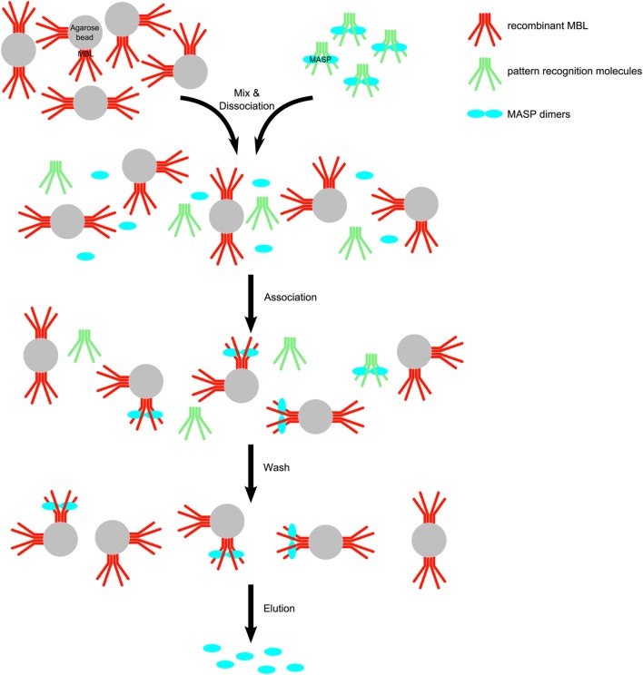
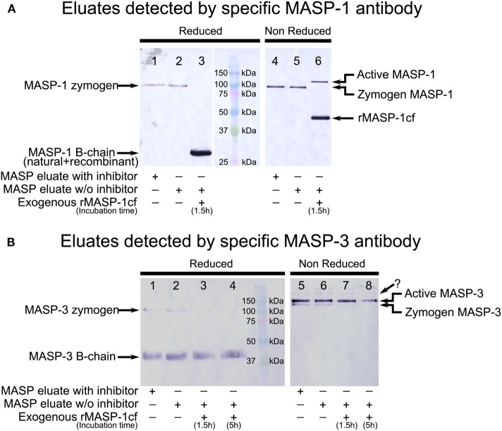
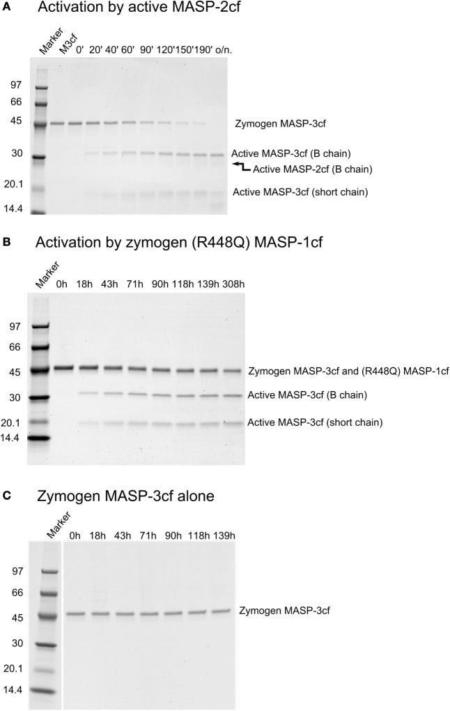
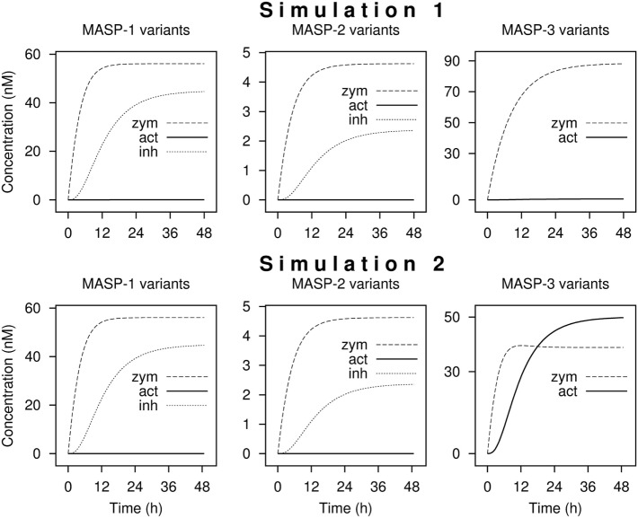
Serine proteases (SPs) are typically synthesized as precursors, termed proenzymes or zymogens, and the fully active form is produced via limited proteolysis by another protease or by autoactivation. The lectin pathway of the complement system is initiated by mannose-binding lectin (MBL)-associated SPs (MASP)-1, and MASP-2, which are known to be present as proenzymes in blood. The third SP of the lectin pathway, MASP-3, was recently shown to be the major activator, and the exclusive "resting blood" activator of profactor D, producing factor D, the initiator protease of the alternative pathway. Because only activated MASP-3 is capable of carrying out this cleavage, it was presumed that a significant fraction of MASP-3 must be present in the active form in resting blood. Here, we aimed to detect active MASP-3 in the blood by a more direct technique and to quantitate the active to zymogen ratio. First, MASPs were partially purified (enriched) from human plasma samples by affinity chromatography using immobilized MBL in the presence of inhibitors. Using this MASP pool, only the zymogen form of MASP-1 was detected by Western blot, whereas over 70% MASP-3 was in an activated form in the same samples. Furthermore, the active to zymogen ratio of MASP-3 showed little individual variation. It is enigmatic how MASP-3, which is not able to autoactivate, is present mostly as an active enzyme, whereas MASP-1, which has a potent autoactivation capability, is predominantly proenzymic in resting blood. In an attempt to explain this phenomenon, we modeled the basal level fluid-phase activation of lectin pathway proteases and their subsequent inactivation by C1 inhibitor and antithrombin using available and newly determined kinetic constants. The model can explain extensive MASP-3 activation only if we assume efficient intracomplex activation of MASP-3 by zymogen MASP-1. On the other hand, the model is in good agreement with the fact that MASP-1 and -2 are predominantly proenzymic and some of them is present in the form of inactive serpin-protease complexes. As an alternative hypothesis, MASP-3 activation by proprotein convertases is also discussed.

Keywords: autoactivation; complement; innate immunity; lectin pathway; proenzyme; reaction kinetics; serine protease.


Figures

Figure 1
Figure 1
Scheme for the purification of mannose-binding lectin (MBL)-associated serine protease (MASPs) from human plasma. Human EDTA plasma was mixed with recombinant human mannan-binding lectin (rMBL)–Sepharose in the presence of high salt (1 M NaCl) causing to dissociate MASP– pattern recognition molecule (PRM) complexes. The ionic strength was decreased by dilution and excess CaCl2 was added to allow complexes to reassociate. MASPs (blue) are distributed between the original PRMs (green) from blood and the immobilized MBL (red). Soluble PRMs and soluble complexes are removed during the wash step. After washing, MASPs bound to rMBL-Sepharose were eluted with a buffer containing EDTA and high salt.
Figure 2
Figure 2
Western blot detection of mannose-binding lectin (MBL)-associated serine protease (MASP)-1 and MASP-3 isolated from normal human plasma in the presence of inhibitors. A pool of MASPs was purified from human plasma as outlined in Figure 1 in the presence of Pefabloc and 4-nitrophenyl 4′-guanidinobenzoate (NPGB) inhibitors. A sample was prepared in the same manner except that the inhibitors were omitted at the last elution step. Samples were analyzed by SDS-PAGE under reducing and non-reducing conditions followed by Western blotting and detected using a MASP-1- or a MASP-3-specific antibody. Both antibodies were developed against the serine protease (SP) domain of the corresponding protein, hence they can detect the whole molecule, or the B chain of the active form. (A) MASP-1 was present as a zymogen in the isolated samples running at about 95 kDa under reducing conditions. When exogenous active recombinant MASP-1cf was added to the inhibitor-free sample, plasma MASP-1 is converted to the active form. The B chains of plasma MASP-1 and MASP-1cf both ran at about 28 kDa under reducing conditions. Under non-reducing conditions, activation of plasma MASP-1 produced a slower-migrating band compared to the zymogen form. MASP-1cf runs at about 45 kDa under non-reducing conditions. (B) MASP-3 was present both in the zymogen and the active forms in the isolated samples. The zymogen ran at about 100 kDa under reducing conditions and the (glycosylated) B-chain of the active form ran at about 40 kDa. Under non-reducing conditions active MASP-3 migrated slower than the zymogen form. The faint band, indicated by a question mark, running above the active form is probably due to non-specific binding. Addition of active recombinant MASP-1cf to the inhibitor-free sample caused the disappearance of the MASP-3 zymogen bands.
Figure 3
Figure 3
Western blot analysis of purified mannose-binding lectin (MBL)-associated serine protease (MASP) pools detected by a MASP-3-specific antibody. MASPs were purified from human plasma as outlined in Figure 1 in the presence of Pefabloc and 4-nitrophenyl 4′-guanidinobenzoate (NPGB). Samples were analyzed by SDS-PAGE under non-reducing conditions followed by Western blotting and detection using a MASP-3-specific antibody. The faint band running above the active form is probably due to non-specific binding of the antibody. Western blots were quantified as described in the Section “Materials and Methods” and the result are listed in Table 1. (A) The analysis of three parallel preparations starting from the same pool of human EDTA plasma. (B) The analysis of samples purified from the plasma of seven individuals. The full blots are provided as Figure S1 in Supplementary Material.
Figure 4
Figure 4
Activation of zymogen mannose-binding lectin (MBL)-associated serine protease (MASP)-3cf. Zymogen MASP-3cf at 2 µM was incubated with active MASP-2cf, or zymogen R448Q MASP-1cf, or alone. Aliquots were removed periodically at time points as indicated. Samples were analyzed by SDS-PAGE under reducing conditions. Molecular weights of the marker proteins in kDa are indicated. Zymogen MASP-3cf runs at about 48 kDa, while the active form gives two bands at about 31 and 17 kDa. Representative gels are shown as examples. (A). Activation by 91 nM active MASP-2cf. The band corresponding to the B chain of MASP-2cf (about 27 kDa) is very faint due to its low concentration. Lane 2 had zymogen MASP-3cf (M3cf) alone. (B) Activation by 1 µM zymogen R448Q MASP-1cf. The band of zymogen R448Q MASP-1cf (about 46 kDa) comigrated wit that of zymogen MASP-3cf. Quantification was carried out as described in the Section “Materials and Methods” and the determined rate constant are listed in Table 2. (C) Zymogen MASP-3cf alone is shown as a control demonstrating that it does not autoactivate or cleaved by any potential contaminant upon prolonged incubation.
Figure 5
Figure 5
Kinetic simulations of the activation of lectin pathway proteases. Time courses of the concentrations of zymogen (zym), active (act), and combined C1-inhibitor and antithrombin bound (inh) forms of MASP proteases from kinetic simulations are shown starting with zero levels of mannose-binding lectin (MBL)-associated serine protease (MASPs). Simulation 1 was performed with measured rate constants. For Simulation 2, the rate constant of the activation of MASP-3 by zymogen MASP-1 was increased by a factor of 200. The full set of kinetic parameters is found in Table S1 in Supplementary Material. The reaction set is depicted in Figure 6 within the boxed area. Steady-state distributions are shown in Table 3.
Figure 6
Figure 6
Possible mechanisms of mannose-binding lectin (MBL)-associated serine protease (MASP)-3 activation. The boxed part depicts activation and inactivation steps involving only lectin pathway proteases. Three forms of MASP-1 (zM1 for zymogen MASP-1, aM1 for active MASP-1, and iM1 for serpin-inhibited MASP-1), three forms of MASP-2 (zM2 for zymogen MASP-2, aM2 for active MASP-2, and iM2 for serpin-inhibited MASP-2), and two forms of MASP-3 (zM3 for zymogen MASP-3 and aM3 for active MASP-3) are shown as separate variants. Red arrows pointing from the enzyme to the substrate represent previously established activation reactions involving MASP-1 and MASP-2. Pink arrows (pointing from the enzyme to the substrate) represent possible activation reactions producing active MASP-3. Cleavage of zymogen MASP-3 by zymogen MASP-1 is highlighted by thick pink arrow (again pointing from the enzyme to the substrate). Inhibition by serpins is indicated by blue T-shaped symbols. Black arrows simply indicate conversion.

References

    1. Merle NS, Church SE, Fremeaux-Bacchi V, Roumenina LT. Complement system part I – molecular mechanisms of activation and regulation. Front Immunol (2015) 6:262.10.3389/fimmu.2015.00262 - DOI - PMC - PubMed
    1. Ricklin D, Hajishengallis G, Yang K, Lambris JD. Complement: a key system for immune surveillance and homeostasis. Nat Immunol (2010) 11:785–97.10.1038/ni.1923 - DOI - PMC - PubMed
    1. Sarma JV, Ward PA. The complement system. Cell Tissue Res (2011) 343:227–35.10.1007/s00441-010-1034-0 - DOI - PMC - PubMed
    1. Dobó J, Pál G, Cervenak L, Gál P. The emerging roles of mannose-binding lectin-associated serine proteases (MASPs) in the lectin pathway of complement and beyond. Immunol Rev (2016) 274:98–111.10.1111/imr.12460 - DOI - PubMed
    1. Nilsson B, Ekdahl K. The tick-over theory revisited: is C3 a contact-activated protein? Immunobiology (2012) 217:1106–10.10.1016/j.imbio.2012.07.008 - DOI - PubMed